
 Türkçe
 Français
 Deutsch

Periodontitis treatment in Turkey involves comprehensive periodontal therapy aimed at eliminating infection, controlling inflammation, and preserving supporting bone and soft tissue structures. Treatment protocols include scaling and root planing, advanced laser applications, and when necessary, surgical intervention performed by qualified periodontists.
Cost of periodontitis treatment in Turkey is determined by disease severity, number of affected teeth, and the need for adjunctive surgical procedures. Compared to many European countries, Turkey offers cost-effective periodontal care while maintaining high clinical standards, modern technology, and internationally accredited dental facilities.
Advanced periodontal surgery options in Turkey include flap surgery, guided tissue regeneration, and bone grafting techniques designed to restore lost periodontal support. These procedures are conducted under strict sterilization protocols and individualized treatment planning to ensure optimal healing and long-term stability.
Recovery process after periodontitis treatment in Turkey depends on oral hygiene compliance, systemic health status, and regular professional follow-up. Post-treatment care focuses on plaque control, antimicrobial support when indicated, and structured maintenance visits to prevent recurrence and protect periodontal health.
| What You Need to Know | Information |
| Definition | Periodontitis is a serious gum disease where inflammation of the gums (gingivitis) progresses and spreads to the bone tissue supporting the tooth, causing permanent damage. If left untreated, it can lead to tooth loss. |
| Progression | Bacteria in dental plaque first cause gum inflammation (gingivitis), which then progresses to reach from the gums to the jawbone, resulting in periodontitis. |
| Risk Factors | – Poor oral hygiene – Smoking and tobacco use – Genetic predisposition – Diabetes – Stress – Immune system diseases – Hormonal changes (pregnancy, menopause) |
| Symptoms | – Gum recession – Persistent bleeding gums – Tooth mobility and shifting – Bad breath – Pain while chewing – Deep pocket formation between the tooth and gum |
| Stages | – Initial Stage: Inflammation and slight recession of the gums are observed. Bone loss has begun. – Moderate Periodontitis: Deep gum pockets form, and bone destruction progresses. Mild tooth mobility may be seen. – Advanced Periodontitis: Severe bone loss and significant tooth mobility are present. The risk of tooth loss is high. |
| Diagnostic Methods | – Measuring pocket depth with a periodontal probe – Evaluation of bone loss with panoramic or periapical X-rays – Clinical examination to determine tooth mobility and inflammation |
| Treatment Methods | – Initial Stage: Tartar removal (scaling) and root surface debridement (curettage), effective oral hygiene training – Moderate Stage: Deep curettage procedures and use of antibacterial agents – Advanced Stage: Surgical treatment (flap operation), bone graft applications, possible tooth extraction |
| Cases Requiring Surgical Intervention | – Deep bone pockets – Severe gum recession causing aesthetic problems – Advanced tissue loss requiring regenerative procedures |
| Post-Treatment Process | After treatment, the patient should maintain oral hygiene and have regular periodontal check-ups and cleanings (usually every 3–6 months). |
| Permanent Effects | Untreated periodontitis can lead to tooth loss, permanent damage to the jawbone, and systemic complications that may affect overall health (e.g., it may increase the risk of cardiovascular diseases). |
| Prevention Methods | – Effective daily oral care (brushing, flossing) – Regular professional cleaning – Healthy diet – Quitting smoking – Keeping systemic diseases under control |
What Is Periodontitis?
Periodontitis is an advanced gum disease resulting from the inflammation and damage of the tissues surrounding the teeth (gums, periodontal ligament, and bone). If left untreated, it can lead to tooth loss. It usually starts when dental plaque is not cleaned for a long time, and the bacterial infection gradually progresses beneath the gumline.
What Are the Symptoms of the Disease and How Can You Monitor Yourself?
The most dangerous aspect of periodontitis is that it often progresses without pain. Since you don’t feel a throbbing pain like with a cavity, you may assume “everything is fine.” However, your body constantly sends you signals. The most common and earliest sign is bleeding. The pink color you see in the sink while brushing, the blood stain left on a firm apple after biting, or blood marks on your pillow in the morning are not normal. Healthy tissue does not bleed, no matter how much you brush.
As the disease progresses, the color and texture of the gums change. While healthy gums are light pink, stippled like orange peel, and tightly hug the teeth, diseased gums become red, purplish, swollen, shiny, and loose. When the gums start to recede, you may notice your teeth appear longer. Besides the aesthetic deterioration, exposed root surfaces lead to sudden sensitivity to heat and cold.
In more advanced stages, due to the loss of supporting tissue, the teeth begin to loosen. You may notice gaps forming between teeth that used to be close together, and feel your teeth move while eating. Bad breath is also a very noticeable sign because the pus and decaying food particles accumulated in deep pockets cause a persistent and stubborn odor. Sometimes, when you press on the gum with your finger, you may even see a whitish, thick fluid (pus) come out from between the tooth and gum.
The main symptoms you should pay attention to are:
- Gum bleeding
- Persistent bad breath
- Swelling in the gums
- Redness in the gums
- Gum recession
- Appearance of longer teeth
- Spaces between teeth
- Tooth mobility
- Difficulty chewing
- Pus discharge from the gum line
What are the causes of this disease and what role does bacterial plaque play?
The origin of the disease is multifactorial—there is not a single culprit, but the leading actor is always bacterial biofilm. Hundreds of types of microorganisms live in our oral cavity. Under normal conditions, they are in balance. However, when oral hygiene is neglected, a sticky, colorless layer called “microbial dental plaque” accumulates on tooth surfaces and at the gumline. This biofilm layer is the primary cause of periodontitis.
This plaque is not just food debris; it is an organized community of bacterial colonies. If not cleaned regularly, it hardens by combining with minerals in saliva and turns into tartar (calculus). Tartar, due to its rough surface, allows more plaque to adhere and mechanically irritates the gums continuously.
But here’s the interesting part: plaque initiates the disease, but the severity of destruction is determined by your immune system. The body wages a war against these bacteria. Defense cells flood the area. Unfortunately, the enzymes released during this battle not only destroy the bacteria but also damage your own tissues. So, periodontitis is not just a bacterial infection—it’s a destructive condition resulting from the host’s inflammatory response. Therefore, treatment does not only aim to remove bacteria but also to control the body’s excessive reaction.
The key factors contributing to disease progression are:
- Poor oral hygiene
- Bacterial plaque accumulation
- Tartar formation
- Rough filling surfaces
- Ill-fitting prostheses
- Genetic predisposition
How do risk factors like smoking and diabetes affect the process?
Some people brush their teeth very little but do not lose them, while others are very meticulous and still face serious issues. This is where “risk factors” come into play. The two major factors that most alter the course of periodontal disease are smoking and diabetes.
Smoking is a disaster for periodontal health. It constricts the capillaries in the gums and reduces blood flow. This has two negative consequences. First, because the gums do not bleed, the patient may not realize the disease exists—we call this a “masked disease.” Second, and more importantly, since tissue nourishment is impaired, defense cells cannot reach the area, and healing capacity collapses. In patients who smoke 10 or more cigarettes per day, bone destruction progresses much faster, and the success rate of treatments is significantly lower compared to non-smokers.
The relationship between diabetes and gum disease is bidirectional. In patients with uncontrolled diabetes (those with high HbA1c levels), blood vessel structure deteriorates, and the immune system weakens. This causes gum infections to progress much more aggressively. Conversely, having a severe infection in the mouth (periodontitis) also makes it harder to regulate blood sugar. Because overall inflammation in the body increases, insulin resistance may be triggered. Therefore, it is vital that diabetic patients pay extra attention to their gum health.
Systemic and environmental factors that worsen the disease include:
- Smoking
- Uncontrolled diabetes
- Obesity
- Stress
- Hormonal changes
- Immunosuppressive medications
- Poor dietary habits
What are the stages of periodontitis and what does the 2017 classification mean?
In 2017, the scientific community and we clinicians adopted an updated classification system to better define the disease and plan accurate treatment. We no longer separate the disease as “chronic” or “aggressive.” Instead, we use a “Staging” and “Grading” system similar to cancer staging. This system tells us both the current severity of the disease and how quickly it might progress in the future.
Staging reflects the extent of damage. Stage I is the earliest phase; bone loss is minimal, and tissue loss is 1–2 mm. If caught at this point, complete reversal is possible. In Stage II, damage reaches a moderate level, and nearly one-third of bone support is lost. Stage III involves significant destruction; pockets have deepened, furcation involvement has occurred, and some teeth may already be lost due to disease. Stage IV is the most advanced stage; many teeth have been lost, the remaining ones are mobile, and chewing function is at risk of total collapse. At this point, we must rehabilitate not just the gums but the entire jaw function.
Grading reflects the “rate” and “risk” of disease progression. Here, the patient’s biological profile plays a role. For example, in a non-smoker without diabetes, the disease progresses slowly (Grade A or B), while in someone who smokes a pack a day or has uncontrolled diabetes, it progresses very rapidly (Grade C). We base our treatment plan on this speed—patients with fast progression are called in for more frequent check-ups and undergo more aggressive treatments.
Criteria used to determine staging include:
- Clinical attachment loss
- Radiographic bone loss
- Number of lost teeth
- Pocket depths
- Tooth mobility
- Furcation involvement
What is the diagnosis process and what examinations are performed in the clinic?
A diagnosis of periodontal disease cannot be made by simply looking in the mouth. It requires a detailed, systematic, and measurement-based examination process. When you sit in the dentist’s chair, the first thing we do is listen to your complaints and general health history. Details like diabetes, heart disease, or smoking are critical clues for us.
The most important part of the clinical exam is the “probing” process. We use a thin, blunt-tipped instrument called a probe that is marked in millimeters. This procedure does not damage your teeth or pierce your gums. Using the probe, we gently insert it between the tooth and gum to measure pocket depth. In a healthy gum, this depth ranges from 1–3 mm. However, measurements of 4 mm or more indicate disease and bone loss. We take measurements from six different points around each tooth to create a map of your mouth.
During this measurement, we also check whether the gums bleed (BOP – Bleeding on Probing). Bleeding is the clearest sign that the disease is active. We also assess tooth mobility, gum recession, and the amount of plaque accumulation.
We always support this clinical data with radiographic examination. With panoramic X-rays or more detailed periapical films, we observe the type (horizontal or vertical) and the amount of bone loss. We determine how close the bone loss is to the root tip. However, X-rays are two-dimensional and do not show everything; that’s why we combine probing measurements with radiography to reach a definitive diagnosis.
Diagnostic methods used include:
- Periodontal probing
- Bleeding index check
- Plaque index measurement
- Mobility test
- Panoramic X-ray
- Periapical X-ray
- Furcation examination
What are the non-surgical treatment methods and what does phase I therapy aim to achieve?
The foundation and first stage of periodontal treatment consists of non-surgical methods we call “Phase I” or “Initial Therapy.” Regardless of the stage of the disease, treatment always starts with this phase. The main goal is to reduce the bacterial load causing the disease, eliminate inflammation, and create a clean surface for the gum to reattach to the tooth. Often, Stage I and Stage II cases can fully recover with just this phase.
The primary procedure we perform during this stage is “scaling,” or tartar removal. Using ultrasonic devices, we remove heavy tartar and plaque on the tooth surface and gumline through vibration. These devices do not damage the enamel; they only remove adhered deposits.
Then comes the more critical procedure—“Root Surface Planing” (Curettage). This is the part where we intervene deeper in the gum pocket, to the root surfaces. It is usually performed under local anesthesia, so the patient feels no pain. Using special hand instruments (curettes), we scrape off infected cementum tissue, deep tartar, and bacterial toxins embedded in the root surface. We smoothen the root surface. A smooth surface is essential because bacteria cannot adhere easily to slippery surfaces, and the healing gum tissue can better adapt to the smooth root.
During this process, the patient receives individualized oral hygiene education. No matter how well we clean, if the patient doesn’t continue care at home using a brush and interdental brush, bacteria will recolonize within 24 hours. In some cases, especially with aggressive infections or systemic illnesses, antibiotic support may also be necessary in addition to mechanical cleaning.
Procedures performed during initial therapy include:
- Tartar cleaning
- Root surface planing
- Polishing
- Oral hygiene education
- Correction of old fillings
- Elimination of plaque-retentive factors
When is surgical treatment necessary and how is a flap operation performed?
We perform initial therapy, wait a few weeks for tissue healing, and conduct a follow-up examination. If there are still bleeding pockets deeper than 5–6 mm or very advanced bone loss (Stage III and IV), we must intervene “visually.” Because it is sometimes impossible to clean the bottom of deep pockets by hand alone. This is when surgical treatment—Phase II—comes into play.
The most common form of periodontal surgery is the “Flap Operation.” In this procedure, performed under local anesthesia, the gum tissue is gently separated from the tooth and bone with a small incision. This allows the dentist to clearly visualize the root surfaces and the erosion craters in the bone. Inflamed tissues (granulation tissue) are completely removed. Root surfaces are flawlessly planed.
Another major advantage of surgical treatment is the opportunity for “regeneration,” or tissue regeneration. If the bone loss is in a suitable form (typically vertical, crater-like defects), bone grafts (bone powder) and membranes (barrier membranes) can be placed in the area. These materials act as a scaffold for the body to regenerate its own bone and support tissues. Safe materials derived synthetically, from animals, or humans are used.
In contrast, when there is excessive gum overgrowth, a procedure called “Gingivectomy” may be used to cut away excess gum tissue and shallow the pockets. In addition, grafts made from tissue taken from your palate to cover gum recessions are part of this surgical phase. At the end of the procedure, the gums are closed with sutures. The healing process usually takes 1–2 weeks, and thanks to modern anesthetics and painkillers, the process is quite comfortable.
Surgical methods used include:
- Flap operation
- Guided tissue regeneration
- Bone graft application
- Membrane application
- Free gingival graft
- Connective tissue graft
- Gingivectomy
- Gingivoplasty
What is the post-treatment care process and does the disease recur?
The most critical—and perhaps most neglected—part of periodontal treatment is the post-treatment phase. Periodontitis, like diabetes or high blood pressure, is a chronic disease. It is not something we can say, “I’m treated, it’s over, it will never come back.” The disease is brought under control and halted, but the risk of recurrence is always present if care is abandoned.
After the active phases of treatment are completed, “Phase III” or “Maintenance Therapy” begins. This is a lifelong partnership. Based on the patient’s risk status (do they smoke, have diabetes, what is their genetic predisposition?), regular check-ups are required every 3, 4, or 6 months. We call these appointments “recall” sessions. More than just a simple cleaning is done in these sessions—each pocket is checked individually, disease reactivation is assessed, and risk analysis is updated.
The biggest share of success actually belongs to the patient. No matter how perfect the surgery is, if the patient does not remove biofilm at home every day, the disease will return. Besides brushing, flossing and especially using interdental brushes in areas with gaps is not optional—it is essential.
Additionally, maintaining systemic health is part of the treatment. Quitting smoking, controlling blood sugar, and maintaining a balanced diet directly impact treatment success. The goal is not just to keep teeth in the mouth, but to improve the patient’s quality of life—to help them eat comfortably and smile confidently. With regular maintenance and dentist supervision, even advanced tooth loss can be prevented, and natural teeth can be preserved for a lifetime.
What should be done for long-term success:
- Regular dental check-ups
- Effective tooth brushing
- Use of interdental brushes
- Use of dental floss
- Quitting smoking
- Blood sugar control
- Healthy nutrition
Before & After Photos


Frequently Asked Questions
How is gingivitis, the early stage of periodontitis, recognized?
Gingivitis presents with gum bleeding, redness, and swelling. At this stage, bone loss has not yet occurred and it can be completely reversed with treatment.
Which systemic diseases are associated with periodontitis?
Periodontitis is directly related to systemic diseases such as diabetes, heart disease, and rheumatoid arthritis. It can lead to chronic inflammation in the body, negatively affecting the course of these diseases.
How does smoking increase the risk of periodontitis?
Smoking reduces oxygen supply to the gums and weakens the immune system, accelerating the development of periodontitis. It also significantly reduces the response to treatment.
Why does the risk of developing periodontitis increase during pregnancy?
Hormonal changes cause gum tissues to become more sensitive. This makes it harder to defend against bacterial plaque, increasing the risk of periodontitis.
What complications occur in the advanced stages of periodontitis?
Advanced periodontitis can lead to tooth mobility, bone loss, and tooth loss. Permanent damage to the jawbone may also occur.
How is deep tartar cleaning performed in the treatment of periodontitis?
With root surface planing and deep curettage, tartar and bacterial plaque are removed. This procedure helps control the infection under the gumline.
How is periodontitis treated surgically?
In advanced stages, the gum flap is lifted to clean infected tissues and restore lost support structures with bone grafts. These surgical procedures are usually performed under local anesthesia.
What is the healing process like after periodontitis treatment?
Healing of the gums begins within 1–2 weeks after treatment. However, full recovery may take months. Regular check-ups and good oral hygiene accelerate the healing process.
Why does bad breath occur in patients with periodontitis?
Sulfur compounds resulting from bacterial infection cause bad breath. Once the infection is controlled through treatment, this problem typically resolves.
What precautions should be taken to prevent recurrence of periodontitis?
Regular dental check-ups, professional cleanings, brushing at least twice a day, and flossing are effective in preventing recurrence of periodontitis.
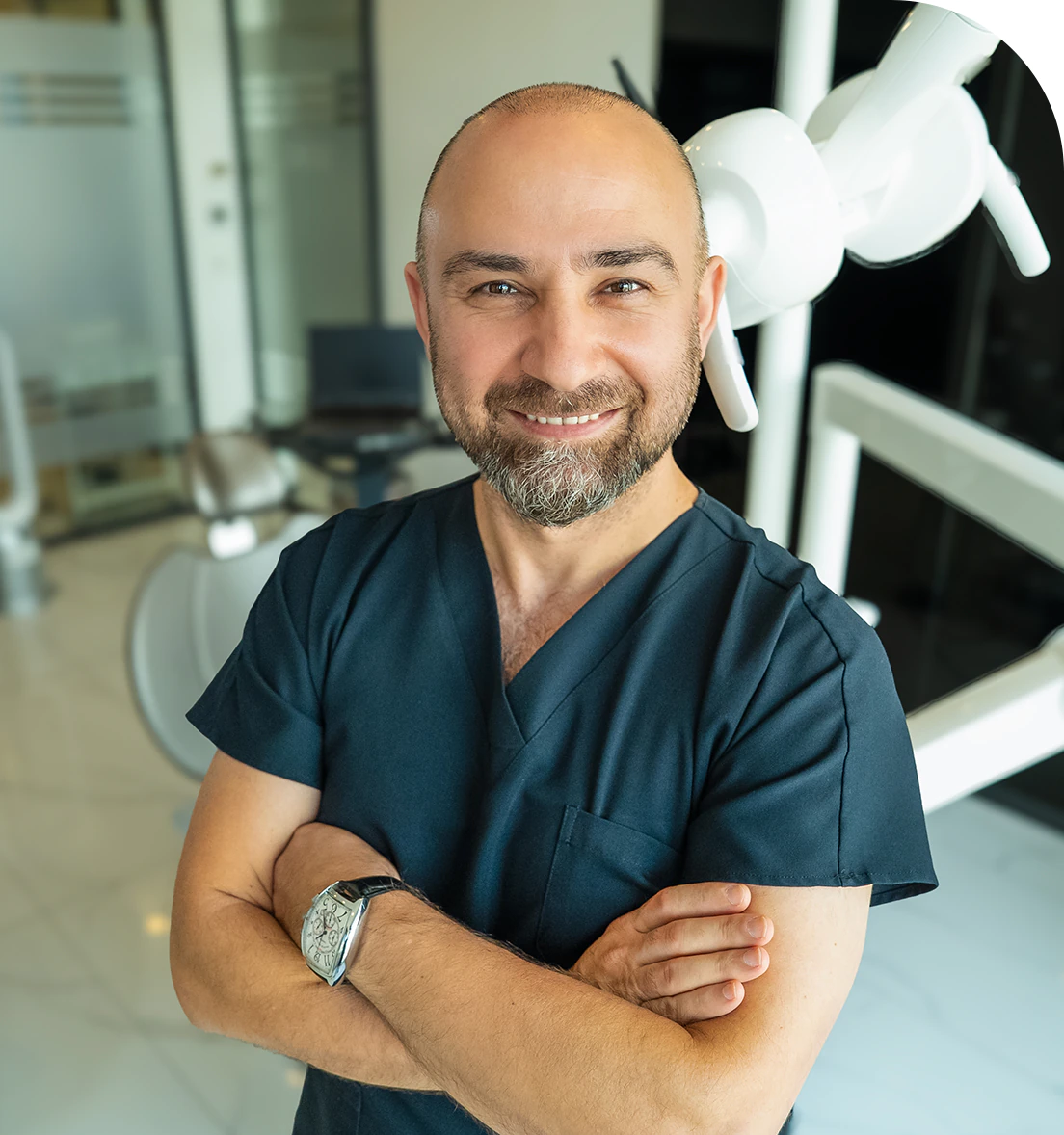
Dr. Dt. Ömer Atar provides care at his private clinic in Istanbul, combining his academic background and many years of clinical experience in dentistry. With a treatment approach that integrates aesthetic and functional solutions, he aims to achieve natural, balanced, and long-lasting results through personalized planning for each patient. Dr. Atar completed his education at the Marmara University Faculty of Dentistry, which he began in 1994, graduating in 2000. He continued his academic work in the Department of Prosthodontics at the same university. In 2007, he earned his PhD degree with his scientific thesis on implants and implant-supported prostheses in patients undergoing distraction osteogenesis. His academic expertise deepened in the fields of metal-free aesthetic systems, implant-supported prostheses, and cosmetic dentistry. Integrating modern technologies and digital dentistry into his practice, Dr. Ömer Atar achieves high success rates in treatments such as smile design, All-on-4/All-on-6 implant treatments, laminate veneers, zirconium crowns, teeth whitening, and gingival aesthetics (gummy smile). As a member of EDAD (Turkish Academy of Aesthetic Dentistry), he embraces a holistic approach focused on modern, functional, and natural outcomes in aesthetic dentistry.


