
 Türkçe
 Français
 Deutsch

Orthognathic surgery in Turkey is a comprehensive surgical procedure performed to correct jaw misalignment and related functional disorders. It aims to improve chewing, speech, breathing, and facial symmetry through precise repositioning of the upper and lower jaws under general anesthesia in accredited medical facilities.
Cost of orthognathic surgery in Turkey is determined by surgical complexity, hospital standards, surgeon expertise, and whether single or double jaw correction is required. Treatment packages often include preoperative diagnostics, hospitalization, anesthesia services, and postoperative follow-up within internationally accredited centers.
Recovery process after orthognathic surgery in Turkey involves a structured postoperative care plan including pain management, nutritional support, and orthodontic adjustments. Initial swelling and mild discomfort are expected, while complete bone healing and stabilization generally occur over several months under specialist supervision.
Advantages of having jaw correction surgery in Turkey include experienced maxillofacial surgeons, advanced imaging technologies, and multidisciplinary treatment planning. Hospitals adhere to strict sterilization and safety protocols, ensuring high clinical standards and predictable functional and aesthetic outcomes for international patients.
| What You Need to Know | Information |
| Definition | Orthognathic surgery is a jaw surgery performed to correct the positions of the jawbones (upper jaw – maxilla and lower jaw – mandible). Skeletal abnormalities in the jaw are corrected both aesthetically and functionally. |
| Purpose | It is used in the treatment of jaw closure disorders (malocclusion), jaw asymmetries, underdeveloped or overdeveloped jaws, facial imbalance, and speech or breathing problems. It is preferred in skeletal disorders that cannot be corrected by orthodontic treatment alone. |
| Who Is It For? | – Individuals with jaws positioned forward or backward from normal – Those with bite issues despite straight teeth – Those with noticeable facial asymmetry or imbalance – Adult (skeletally mature) patients |
| Preoperative Preparation | Orthodontic treatment (with braces) is applied to the patient. This process usually takes 12–18 months. After orthodontic preparation, the jaws are made suitable for surgery. |
| Procedure | Performed under general anesthesia. Jawbones are cut, repositioned, and fixed with titanium plates and screws. The surgery typically lasts between 2 to 4 hours. |
| Postoperative Period | Swelling, pain, and difficulty swallowing may occur in the first few days. A liquid and soft diet is recommended. Return to normal life is usually within 2–3 weeks. Complete recovery can take 6–12 months. |
| Advantages | – Improvement in chewing, speaking, and breathing functions – Noticeable enhancement in facial aesthetics – A permanent and profound solution – Increased psychological and social self-confidence |
| Risks and Complications | – Infection – Bleeding – Temporary or permanent nerve damage (especially numbness in the lower lip) – Temporomandibular joint (TMJ) problems – Difficulty in the postoperative adaptation period |
| Follow-up and Monitoring | Postoperative orthodontic treatment continues for a while. In addition, regular surgical and orthodontic check-ups are required. |
| Alternatives | Since orthognathic surgery offers an effective solution for skeletal problems, there are generally no alternative treatments in cases of severe jaw discrepancies. However, in mild cases, orthodontic treatment alone may be sufficient. |
What Is Orthognathic Surgery?
Orthognathic surgery is a corrective jaw surgery performed to treat congenital or acquired deformities in the shape and position of the jawbones. It is applied in cases where the lower or upper jaw is positioned forward, backward, crosswise, or asymmetrically. In addition to improving facial aesthetics, it also resolves functional problems such as chewing, speaking, and breathing. It is planned in conjunction with orthodontic treatment and usually implemented with a multidisciplinary approach.
Why Is Orthodontic Treatment Alone Insufficient to Solve These Problems?
One of the most frequently asked questions in the clinic is, “Doctor, can’t we fix this just with braces or clear aligners without surgery?” We wish the answer could always be yes, but biology and physics impose certain limits. In professional terms, we refer to this as the “Envelope of Discrepancy.” You can think of it like the foundation and walls of a house—if the foundation is crooked, simply fixing the wallpaper won’t correct the building.
Teeth can be moved within a certain limit inside the jawbone. If the issue is only the alignment of the teeth, orthodontic treatment alone works wonders. But if the problem lies in the supporting bone structure—the foundation—then it is impossible to achieve an ideal bite just by tilting or moving the teeth. If the skeletal discrepancy exceeds a certain millimeter threshold, trying to force the teeth into position does more harm than good. Such force can push tooth roots out of the bone, cause gum recession, and lead to root resorption.
This is when we decide on orthognathic surgery: when the skeletal defect exceeds the safe limits for tooth movement, surgery becomes inevitable. Our goal is not to push teeth into risky zones, but to move the bones into their ideal positions, achieving both aesthetics and lasting health.
How Does the Surgical Planning Process Work and What Technology Is Used?
The success of this treatment begins long before the operating table—at the computer, with detailed planning. In the past, estimates were made using X-rays and plaster models. Today, technology allows us to reduce error margins to nearly zero. The process begins with creating a digital twin of the patient’s face and skeletal structure.
Using Computed Tomography (CT) or Cone Beam Computed Tomography (CBCT), we create a three-dimensional image of the patient’s skull. These images allow us to study the bone structure, tooth roots, nerve pathways, and airways in detail.
Then, as the surgeon and orthodontist, we sit at the computer and perform a virtual surgery. We calculate with millimetric precision how much to move the upper jaw forward, how much to set the lower jaw back, or at what angle to rotate to correct asymmetry. This virtual planning allows us to anticipate any possible scenarios during surgery. Additionally, surgical guide plates tailored to the patient are produced from this data. These guides allow us to transfer the planned movements precisely to the bone during surgery. So, when we enter surgery, every step is mathematically defined, and there’s no room for surprises.
Technologies used in the planning process include:
- 3D scanners
- Cone beam computed tomography
- Virtual surgical simulation software
- 3D printers
- Digital photo analysis tools
- Facial scanning systems
How Is Facial Aesthetics and Soft Tissue Change Targeted?
Although orthognathic surgery is primarily associated with correcting bones, for most patients, the biggest motivation is the change they see in the mirror. Therefore, we don’t just focus on the bones and teeth during planning—we also assess how the soft tissues of the face (lips, nose, cheeks, and chin) will respond to the changes.
In our clinical evaluation, we analyze facial profile balance using what’s called “cephalometric analysis.” For example, when we move the upper jaw forward, we anticipate a slight lift in the nose tip or increased lip support that may enhance fullness. Similarly, when we move the lower jaw back, we assess how the appearance of the chin and neck area may change. Our goal is not only to restore proper chewing but also to achieve an aesthetically balanced facial profile.
At this point, aligning the patient’s expectations with the outcomes of the surgery is crucial. Sometimes the “ideal” skeletal position may not deliver the best aesthetic result. In such cases, we may make minor revisions to the plan, prioritizing aesthetic gains. Ultimately, our goal is not to produce a face that looks “operated on,” but one that is natural, balanced, and well-suited to the individual.
Why Is Collaboration Between the Orthodontist and Surgeon Critical?
The orthognathic surgery process is not managed by a single doctor—it is a team effort. The two main players on this team are the Orthodontist and the Maxillofacial Surgeon (Oral and Maxillofacial Surgeon). The success of the treatment depends directly on the communication and synchronization between these two specialties. The process typically consists of three phases, with responsibility shifting in each phase.
In the first phase—the preparation stage—the orthodontist is in charge. They prepare the teeth for surgery. On the day of surgery—the second phase—the surgeon takes over and moves the bones into the planned position. In the final phase—postoperative finishing—the orthodontist returns to make final adjustments.
If there is a lack of coordination between these two experts, the outcome can be disappointing. For example, if the orthodontist doesn’t prepare the teeth according to the surgical plan, the surgeon may be unable to position the jaws correctly during surgery. Or if the surgeon deviates from the plan, the post-surgery bite may be misaligned. That’s why we hold regular meetings throughout treatment, work together on 3D models, and jointly approve every step. The patient is at the center of this process and is informed at every stage, while behind the scenes, there’s a continuous technical collaboration.
Why Is Pre-Surgical Orthodontic Treatment Necessary?
The part patients often find hardest to understand—and most psychologically challenging—is the “pre-surgical braces” phase. Understandably, they ask, “If I’m getting surgery anyway, why do I need braces for months beforehand?” The answer lies in the body’s natural defense mechanism, known as “compensation.”
In individuals with skeletal defects, the teeth shift over the years to mask the defect by moving into unnatural positions. For example, in someone with a protruded lower jaw, the upper teeth may angle forward and the lower teeth tilt backward to close the gap. We call this “dental camouflage.” If we perform surgery while the teeth are still in this compensated position, they will clash, and we won’t be able to move the jaws adequately.
Therefore, we conduct about 12–18 months of orthodontic preparation before surgery. This process is called “decompensation.” Our goal is to return the teeth to their true and ideal angles relative to the bone. During this phase, an interesting phenomenon occurs: as the teeth straighten, the jaw discrepancy on the face temporarily becomes more noticeable, and the bite worsens. While this may be concerning for patients, it’s a great sign for us—it means the dental camouflage is gone and the space needed for surgical correction has opened up. This phase requires patience, but a strong foundation is essential for an excellent result.
Procedures performed during this period include:
- Correction of tooth crowding
- Aligning dental arches
- Adjusting tooth inclinations
- Placement of surgical archwires
- Taking records and impressions
What Happens on Surgery Day and What Surgical Techniques Are Used?
Once preparations are complete and the teeth have been brought into the desired position, the day of actual transformation arrives. Orthognathic surgery is performed under general anesthesia in a fully equipped hospital setting. The biggest fear among our patients is often whether there will be any scars on their face. I can confidently say that nearly all of these surgeries are performed from inside the mouth (intraoral). That means no visible incision marks will appear on the facial skin.
The most commonly performed procedures are known as “Le Fort I” and “Bilateral Sagittal Split Osteotomy” (BSSO). Although their names may sound complex, their logic is quite straightforward. For the upper jaw, with the Le Fort I technique, an incision is made along a line beneath the base of the nose, and the upper jaw is released. For the lower jaw, the BSSO technique involves a controlled separation at the back portion of the jawbone.
The surgeon moves the bones forward, backward, upward, or downward according to the coordinates determined in the 3D planning. If there is asymmetry, the midline is corrected. Once the bones are positioned in their new place, they are fixed using titanium plates and screws. These plates and screws typically remain in place for life and do not cause any discomfort, are not felt externally, and do not trigger metal detectors at airports.
What Can Patients Expect During the Healing Process?
The postoperative period is when patients need the most physical and emotional support. The hospital stay is usually 1 or 2 days. It is normal to feel tightness in the face and have the jaws banded with elastics upon waking. In the first few days, swelling (edema) rather than pain is predominant. Swelling generally peaks on the third day and then begins to subside rapidly. Applying ice during this period is our greatest aid.
The first month is a “critical healing” period. During this time, as the bones are still in the process of healing, chewing forces must be completely avoided. Patients are advised to eat only liquid or puréed foods during this period. Though adjusting to a new diet can be difficult, it is temporary. After approximately four weeks, patients can transition to soft foods, and as bone healing is confirmed radiographically, a normal diet is gradually resumed.
Recommended foods during the nutrition period include:
- Milk with protein powder
- Blended soups
- Fruit juices
- Yogurt and ayran
- Pulp-free compotes
- Pudding-like desserts
- Vegetable purées
- Meat broth
- Kefir
Why Does Orthodontic Treatment Continue After Surgery?
The end of surgery does not mean the end of treatment. After the surgical procedure, a “postoperative orthodontic treatment” phase lasting approximately 6 to 12 months begins. This phase is similar to the fine finishing work of a building after the main structure is completed. Surgery has corrected the bones significantly, but final touches are needed for the upper and lower teeth to interlock like a zipper.
Elastics and braces used during this phase perfect the relationship between the teeth. Another critical role of this stage is to ensure “stability.” As the muscles and soft tissues try to adapt to the new skeletal structure, maintaining tooth position prevents relapse. Once braces are fully removed, the patient completes treatment both aesthetically and functionally and enters the retention phase with stabilization devices.
What Is the Impact on Patients’ Quality of Life and Psychology?
The gains achieved at the end of this long and challenging journey truly mark a turning point in the patient’s life. We’re not only talking about superficial benefits like “looking better.” Being able to bite into an apple with the front teeth, eating a sandwich without difficulty, or pronouncing words correctly dramatically improves daily comfort.
Especially in young adulthood, correcting prominent facial deformities leads to a burst of self-confidence. Being able to smile freely in social settings, not hiding the face in photos, or not stressing about finding a flattering angle positively affects social interactions. Studies have shown that patients who undergo orthognathic surgery become much happier and more confident individuals psychologically and socially after treatment. Most patients say at the end of the process, “It was a tough road, but it was definitely worth it.”
Can Jaw Joint Problems and Pain Improve?
The Temporomandibular Joint (TMJ), or jaw joint, is one of the most complex structures in the human body. In patients with skeletal deformities, malocclusion causes the joint and masticatory muscles to be exposed to unbalanced loads for years. Over time, this can lead to joint noises (clicking), pain, and limited mouth opening.
Orthognathic surgery aims to eliminate these unbalanced loads. By correcting the bones, muscle function and the forces on the joint normalize. However, after surgery, the joint must adapt to its new position. This is called “condylar remodeling”—the joint head biologically renews itself to fit into its new socket.
In most cases, joint issues decrease or completely resolve after surgery. However, in some cases—especially where the lower jaw is significantly advanced or where there is existing joint damage—careful management of the process is essential. That’s why we monitor joint health before and after surgery and implement additional treatment protocols if needed.
Does It Affect the Airway and Sleep Problems?
One of the least known but most vital health benefits of orthognathic surgery is its effect on the airway. Especially in Class II patients with a retruded lower jaw, the tongue base shifts backward and narrows the airway. This sets the stage for snoring, poor sleep quality, and even serious conditions like Obstructive Sleep Apnea.
Surgeries that bring the jaws forward mechanically widen the airway. This increases the patient’s breathing capacity, improves sleep quality, and enhances oxygenation of the body. On the contrary, in Class III cases where the lower jaw must be moved backward, there is a risk of airway narrowing. That’s why airway analysis during the planning phase is non-negotiable for us. If the airway is at risk, we revise the surgical plan accordingly—because breathing is always a higher priority than aesthetics.
What Should Be Considered for a Successful Outcome?
Success in orthognathic surgery cannot be measured by a single criterion. True success is a combination of a healthy-functioning jaw joint, balanced facial aesthetics, ideal bite closure, a clear airway, and a patient who is happy with their reflection.
To ensure long-term stability of the treatment, the following must be considered:
- Working with an expert team
- Thorough preoperative preparation
- Technologically supported surgical planning
- Patient compliance during the healing period
- Not skipping postoperative check-ups
- Adherence to retention therapy
Before & After Photos


Frequently Asked Questions
In Which Cases Is Orthognathic Surgery Performed?
Orthognathic surgery is performed in cases of jaw discrepancies, severe crowding, or skeletal deformities that cause facial asymmetry. It aims to correct both aesthetic and functional issues.
At What Age Can Orthognathic Surgery Be Performed?
It can be performed after the age of 18, once bone development is complete. It is more suitable during young adulthood for better recovery and longer-lasting results.
Which Orthodontic Treatments Are Combined with Orthognathic Surgery?
It is usually planned in conjunction with fixed orthodontic treatment. Braces are required before and after surgery to bring the teeth into ideal positions, and the entire process can last several years.
What Is the Recovery Process After Orthognathic Surgery?
Swelling and sensitivity may occur during the first 1–2 weeks after surgery. A soft diet is recommended. Full recovery can take up to 6 months, although return to daily life is usually possible within the first month.
What Are the Risks of Orthognathic Surgery?
Risks include bleeding, infection, nerve damage (especially numbness in the lips), and limited jaw movement. These risks can be minimized with experienced surgeons.
How Does Orthognathic Surgery Affect Facial Aesthetics?
As jaw positions are corrected, facial symmetry, profile appearance, and smile aesthetics significantly improve. Patients often gain a more balanced and youthful facial expression.
Does Speech and Chewing Improve After Orthognathic Surgery?
As the bite is corrected, chewing strength increases and speech becomes clearer. There are significant functional gains, especially in severe malocclusion cases.
Is Orthognathic Surgery Covered by Public Health Insurance (SGK)?
Surgeries performed due to functional impairments are generally covered by SGK. However, procedures performed solely for aesthetic purposes may not be included.
What Should the Diet Be Like After Orthognathic Surgery?
In the first week, liquid and puréed foods are recommended. From the second week, patients transition to soft foods. Solid foods are typically introduced within 4–6 weeks under the doctor’s supervision.
Do Plates or Screws Remain in the Jaw After Orthognathic Surgery?
Yes, titanium plates and screws used to fix the jawbones usually remain permanently. Since they are biocompatible, removal is not necessary.
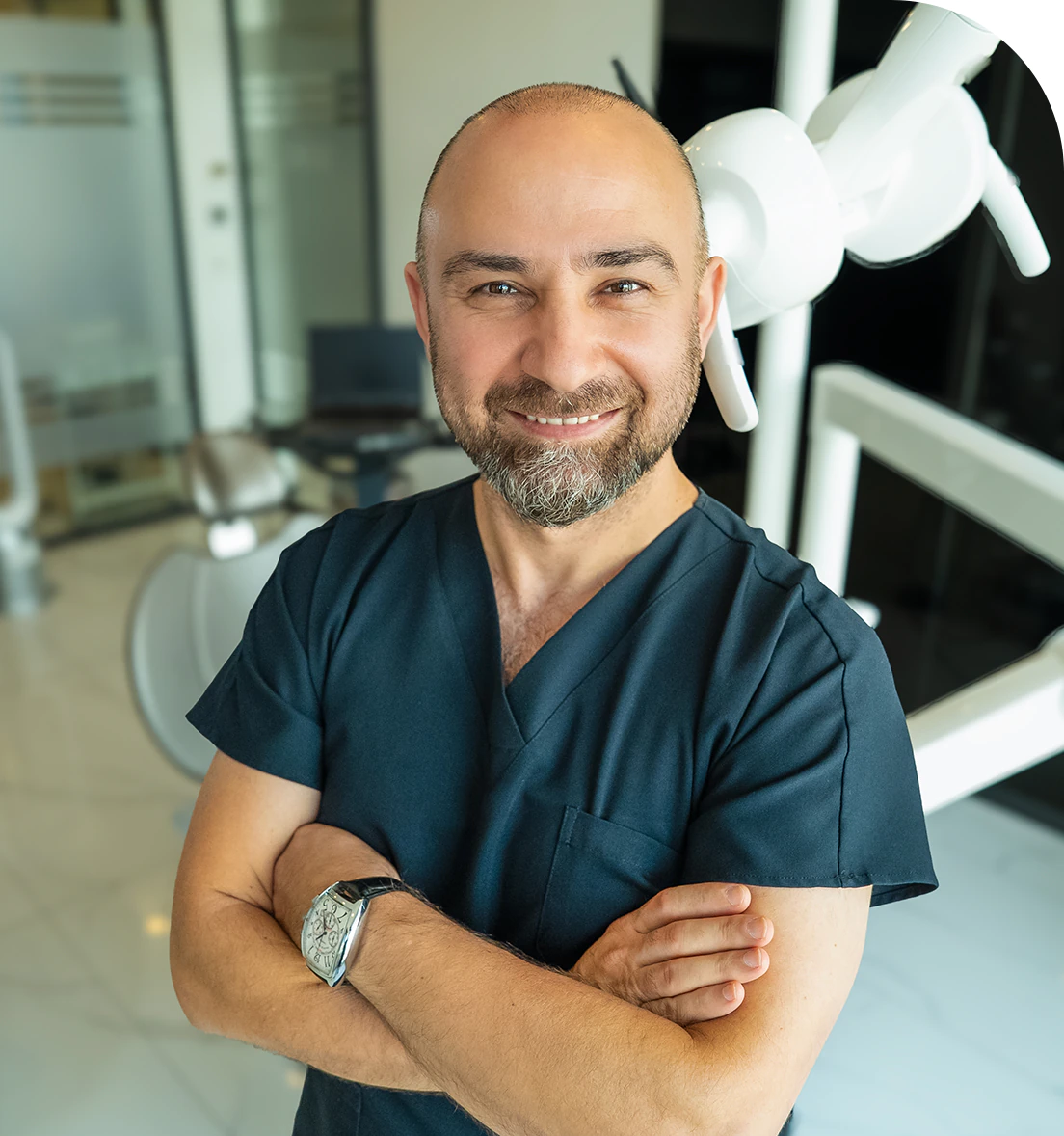
Dr. Dt. Ömer Atar provides care at his private clinic in Istanbul, combining his academic background and many years of clinical experience in dentistry. With a treatment approach that integrates aesthetic and functional solutions, he aims to achieve natural, balanced, and long-lasting results through personalized planning for each patient. Dr. Atar completed his education at the Marmara University Faculty of Dentistry, which he began in 1994, graduating in 2000. He continued his academic work in the Department of Prosthodontics at the same university. In 2007, he earned his PhD degree with his scientific thesis on implants and implant-supported prostheses in patients undergoing distraction osteogenesis. His academic expertise deepened in the fields of metal-free aesthetic systems, implant-supported prostheses, and cosmetic dentistry. Integrating modern technologies and digital dentistry into his practice, Dr. Ömer Atar achieves high success rates in treatments such as smile design, All-on-4/All-on-6 implant treatments, laminate veneers, zirconium crowns, teeth whitening, and gingival aesthetics (gummy smile). As a member of EDAD (Turkish Academy of Aesthetic Dentistry), he embraces a holistic approach focused on modern, functional, and natural outcomes in aesthetic dentistry.


