
 Türkçe
 Français
 Deutsch

There are four main types of teeth in the human mouth, classified according to their functional roles and morphological structures: incisors for biting food, canines for tearing, premolars for crushing, and molars with powerful grinding capacity. Within the biological life cycle, these structures are examined in two main stages: the temporary baby teeth in infancy and the permanent adult teeth in adulthood. Each group of teeth fulfills a specific mechanical function at the beginning of the digestive system while also playing a critical role in speech phonetics, temporomandibular joint health, and facial aesthetics.
What Are the Main Types of Teeth in the Mouth and How Do Their Functions Differ?
We can roughly compare the teeth in our oral cavity to a toolbox. Just as you can’t tighten a screw with a hammer or drive a nail with a screwdriver, your teeth are specialized for different functions. This specialization is the result of millions of years of evolutionary processes and our dietary habits. Essentially, we divide our teeth into four main categories based on their position and shape.
The basis of this categorization lies in “functionality.” The teeth located at the front of the mouth are sharper and thinner, while as you move toward the back, the teeth become broader in surface and stronger in root structure. The reason for this lies in the masseter muscle, which allows us to close our jaws. As one of the strongest muscles in the human body, it can generate pressure in the rear regions of the mouth reaching hundreds of kilograms when the jaw is closed. To withstand this immense force and prepare food for digestion, the back teeth must be as solid as millstones.
Understanding this system helps us grasp why some teeth decay faster or why the face shape changes when certain teeth are extracted. It also sheds light on the origin of jaw joint problems, which often stem from disharmony among these tooth groups, and can change your perspective on treatment approaches. Now let’s explore these groups and their life cycles from the very beginning—starting with infancy.
What Symptoms Are Observed When Baby Teeth Erupt?
Humans have two sets of teeth throughout their lifetime. The first is the primary dentition, commonly known as baby teeth. Typically, the first baby teeth begin to appear between the 6th and 9th months of a baby’s life. However, this is not a strict rule—biology differs from mathematics, and every baby follows their own unique developmental timeline. Some babies may start teething as early as the 3rd month, while in others, the first tooth may not appear until their first birthday. This situation depends on genetic factors and is generally nothing to worry about.
The teething process can be a bit challenging for both the baby and the parents. The gums stretch due to the pressure from the emerging tooth, causing mild inflammation in the area. A baby’s tendency to put everything into their mouth during this time is actually an effort to soothe their itchy and tense gums.
The most common physiological symptoms parents may observe during this process include:
- Increased drooling
- Restlessness and bouts of crying
- Disrupted sleep patterns
- Loss of appetite
- Desire to bite hands
- Redness in the gums
- Mild cough
- Redness in the cheeks
However, it is very important to note the following. In society, beliefs such as “the fever is due to teething” or “the diarrhea is because of the teeth” are quite common. Scientific data shows that teething does not directly cause high fever (38°C and above) or severe diarrhea. During this period, the immune system may temporarily weaken, making babies more susceptible to infections. If your baby has a persistent fever or diarrhea, the safest course is to consult a pediatrician rather than attributing it solely to teething. For discomfort caused by teething, cold (not frozen) teething rings or gels recommended by your doctor can be excellent remedies.
Why Is the Six-Year Molar Considered a “Hidden Treasure”?
One of the most critical milestones in children’s oral and dental health journey occurs around the age of six. At this time, a new tooth quietly erupts at the back of the mouth, right behind the baby molars. This tooth is the First Permanent Molar, also known as the “Six-Year Molar.”
The reason this tooth is referred to as a “hidden treasure” and why it poses significant clinical risk lies in the way it erupts. Usually, a baby tooth loosens and falls out to make space for a new tooth. However, the six-year molar does not replace any baby tooth—it erupts directly from the empty area behind the jawbone. For this reason, parents often mistake it for a baby tooth as well. Thinking “it will fall out anyway,” early cavities in this tooth are frequently neglected. Yet this tooth is the keystone of the chewing system and is meant to stay in the mouth for a lifetime.
Premature loss of the six-year molar can create a domino effect. The chewing center becomes disrupted, other teeth shift into the empty space, and serious crowding may develop, requiring complex orthodontic treatment in the future. That’s why it is vital for every six-year-old child to be examined by a dentist.
To protect these teeth, dentists often apply the following preventive measures:
- Fissure sealant applications
- Topical fluoride treatments
- Toothbrushing education
- Dietary regulation
Fissure sealants are painless and highly effective protective shields that fill in the deep grooves of the chewing surfaces, preventing food particles from accumulating. This simple precaution is one of the best investments you can make for your child’s future dental health.
What Role Do Front Teeth Play in Aesthetics and Smile Design?
The front teeth (incisors), which act as the showcase of our mouth, play a much larger role in our social lives than we might think. When meeting someone, after the eyes, the smile is often the next focal point. The anterior teeth—incisors and canines—not only help bite into an apple but also support the lips to prevent facial collapse and play a key role in the pronunciation of words (phonation).
A beautiful smile is not just about having white teeth. In aesthetic dentistry, “white aesthetics” (teeth) and “pink aesthetics” (gums) are evaluated as a whole. In an ideal smile, all the upper front teeth should be visible, and about 1–2 mm of the gumline should also be perceptible. However, in some cases, the gums appear more than they should—this is known as a “gummy smile.” Conversely, if the teeth are hidden under the lips, the person may appear older than they are.
Today, there are many technological solutions available to address aesthetic issues in the front teeth. If the teeth only have discoloration or slight shape irregularities, a technique called “bonding” can be used to redesign the shape of the tooth with special filling materials without causing any damage to the tooth. For more permanent and dramatic changes, porcelain laminate veneers (also known as leaf porcelain) are preferred.
Common situations where aesthetic intervention may be required for front teeth include:
- Gaps between teeth
- Permanent discoloration
- Fractures or cracks
- Mild crowding
- Size discrepancies
- Worn tooth surfaces
Remember, an aesthetic smile design is highly personalized. A tooth shape that looks great on someone else may not suit your facial structure or lip contour. Your dentist will plan the most suitable smile for you by calculating your facial golden proportions.
What Should Be Done for a Broken Front Tooth?
Due to their location, the front teeth are the most exposed to trauma. A child who falls while running in the school corridor, a teenager in a bike accident, or an adult who gets hit during sports—all face the common nightmare of a fractured front tooth. In such cases, staying calm and taking the right steps can save the life of the tooth.
In cases of fracture, the “golden hour” rule applies. It is critical to reach the dentist within the first 30 to 60 minutes after the incident. If your tooth has broken and you can find the fragment, do so. The broken piece can be reattached using special adhesives (bonding agents) and function just like your natural tooth.
In more severe cases, the tooth may be completely dislodged along with its root (avulsion). In such situations, your initial intervention can make all the difference. When you find the tooth, never hold it by the root. The root surface contains living cells that are essential for reattachment and must not be damaged. Hold the tooth by the white crown part. If it’s dirty, rinse it gently under running water for about 10 seconds without rubbing.
The most appropriate mediums for transporting the tooth are:
- Cold milk
- The patient’s own saliva
- Physiological saline solution
- Inside the cheek
Never wrap the tooth in tissue or store it in a dry container—dried root cells die within minutes. Once you reach the dentist, a treatment plan is made depending on the depth of the fracture. If the fracture hasn’t reached the nerve (pulp), it can be resolved with aesthetic fillings. If the nerve is exposed, root canal treatment may be required. In modern dentistry, the priority is always to keep the natural tooth in the mouth.
Why Do Canines Guide Jaw Movements?
When you look in the mirror, you’ll notice those sharp, strong teeth located at the corners right next to the incisors. These are the canines, also known as cuspids. Commonly referred to as “eye teeth,” their roots are so long that they can extend nearly below the eye socket. This long and sturdy root structure makes them the most resilient teeth in the mouth.
Canines are not only important for tearing and aesthetics; they are also the “navigators” of jaw movements. When we slide our jaw to the right or left, the lower and upper canines come into contact and guide the jaw’s direction. This contact causes the back molars to separate and prevents them from grinding against each other. This mechanism, known as “canine guidance,” acts as a safety system for the chewing function. If your canines are not in the proper position or are worn down, this protection disappears, and your back teeth start clashing during side movements. Over time, this can lead to cracks in teeth, broken fillings, and most importantly, joint pain.
However, due to difficulties in the eruption path, canines may sometimes remain impacted in the palate or lip-side of the jaw. This situation typically becomes detectable around the ages of 11–12.
The risks associated with impacted canines include:
- Resorption (erosion) of neighboring tooth roots
- Cyst formation
- Disruption of dental alignment
- Risk of infection
In such cases, collaboration between an orthodontist and an oral surgeon may be necessary to surgically expose the impacted tooth and bring it into position using orthodontic traction. This treatment requires patience, but considering the functional importance of the tooth, it is undoubtedly worth the effort.
What Is the Relationship Between Back Teeth and Chewing Force?
When we move to the posterior region of the mouth—the “kitchen” zone—the structure of the teeth changes entirely. Premolars and molars have broad chewing surfaces designed for grinding, crushing, and preparing food for digestion. This is where strength and durability matter most.
In treatments involving this area, unlike the front teeth, mechanical durability takes precedence over aesthetics. That’s because the amount of force applied to these teeth during chewing is tremendous. If a back tooth is decayed or requires a root canal, the material chosen by your dentist must be able to withstand this load. In the past, only gray-colored amalgam fillings were used in this area. Today, thanks to advancing technology, reinforced composites or porcelain inlays/onlays can offer both aesthetic appeal and rock-solid strength.
The most critical factor in back tooth restorations is “occlusion,” or how the teeth come together. If a filling or crown is even slightly too high, chewing forces will concentrate on that point. This can cause sensitivity, pain, and even fractures extending to the root.
Key points for protecting back teeth include:
- Avoiding hard, shelled foods
- Using a night guard if you have a habit of clenching or grinding
- Flossing to prevent interproximal decay
- Replacing missing teeth in a timely manner
Should You Choose an Implant or a Bridge for Tooth Loss?
Loss of a back tooth is sometimes neglected with the thought, “It doesn’t show anyway.” But this is a big mistake. Losing even one molar can reduce chewing efficiency by 30–40%. Incomplete digestion in the mouth can trigger stomach problems. More importantly, nature abhors a vacuum. The neighboring teeth begin to tip into the empty space, and the opposing tooth starts to grow into it. This domino effect can lead to the collapse of the entire dental structure within a few years.
There are two main modern solutions for replacing missing teeth: implants and bridges. Both have their advantages and disadvantages. However, in modern dentistry, implants are more favorable due to their ability to preserve biological tissues.
Key features of implant treatment include:
- No need to touch neighboring healthy teeth
- Prevents jawbone resorption
- Provides the closest sensation to natural chewing
- Offers a long-lasting solution
- Maintenance is like that of a natural tooth
Implants are artificial roots made of titanium. After being placed into the jawbone, they are left to integrate (osseointegrate) with the bone. This process is a biological marvel, and titanium becomes part of the bone.
Features of bridge treatment include:
- Faster to complete
- No surgical procedure required
- Lower cost compared to implants
- Requires trimming of supporting teeth
Your dentist will evaluate your bone structure, general health (diabetes, osteoporosis, etc.), and expectations to map out the best solution. However, remember that replacing a lost tooth is not just about filling the space—it’s about restoring the health of the entire system.
Should All Types of Wisdom Teeth Always Be Extracted?
One of the most debated topics in dentistry—and among the most feared by patients—is wisdom teeth (third molars). These teeth usually attempt to erupt between the ages of 17 and 25 and are actually a remnant of our evolutionary heritage. Our ancestors, who consumed tough meats and fibrous root vegetables, needed wide jaws and extra grinding teeth. However, as our diets have changed and become softer and more refined over time, our jaw structures have also shrunk. The result? There’s often no room left for wisdom teeth.
This lack of space causes wisdom teeth to frequently remain impacted or partially impacted. Partially erupted teeth form a pocket between the gum covering and the tooth itself. Food particles trapped in this pocket cannot be cleaned and create a perfect breeding ground for bacteria. This leads to an infection known as “pericoronitis,” characterized by severe pain, limited mouth opening, and facial swelling.
However, not every wisdom tooth is destined for extraction. If a wisdom tooth has erupted at the correct angle, is brushable, and has a functional opposing tooth, there is no harm in keeping it.
Situations that require wisdom tooth extraction include:
- Recurring infections
- Cavities that cannot be restored due to positioning
- Cyst or tumor formation
- Pressure or damage to the neighboring tooth
- Cheek biting issues
- Need for orthodontic treatment
The condition of wisdom teeth is generally monitored with panoramic X-rays. Even if they are asymptomatic, regular checkups are essential to detect possible cyst formation around impacted teeth.
What Types of Radiographic Dental Images Are Used for Diagnosis?
One of the first things a dentist does is take an X-ray. Some patients may wonder, “The problem is visible—why do we need a film?” But an intraoral exam only shows us the tip of the iceberg. The visible part of the tooth (the crown) accounts for only one-third of its structure. The roots, the bone surrounding them, nerve canals, and potential pathologies are hidden deeper within.
It is impossible to apply the correct treatment without an accurate diagnosis. That’s why radiology is the “eyes” of dentistry. Some patients may hesitate due to fear of radiation; however, today’s digital radiographic devices emit very low radiation doses, and the diagnostic benefits far outweigh the risks.
The most common imaging methods used in clinical practice include:
- Periapical X-rays
- Panoramic X-rays
- Dental tomography (CBCT)
- Cephalometric X-rays
Periapical films show a single tooth and its root in detail and are used for procedures like root canal treatment or detecting deep decay. Panoramic films, on the other hand, offer a comprehensive map of the entire jaw, including the joints, sinuses, and impacted teeth—all in one image.
Dental tomography is the pinnacle of technology. It provides 3D imaging. It is indispensable for procedures such as measuring bone thickness down to the millimeter for implants, determining the proximity of an impacted tooth to a nerve, or mapping the boundaries of cysts. Thanks to tomography, your dentist doesn’t have to work “blind,” encounters fewer surprises, and can plan your treatment with maximum safety.
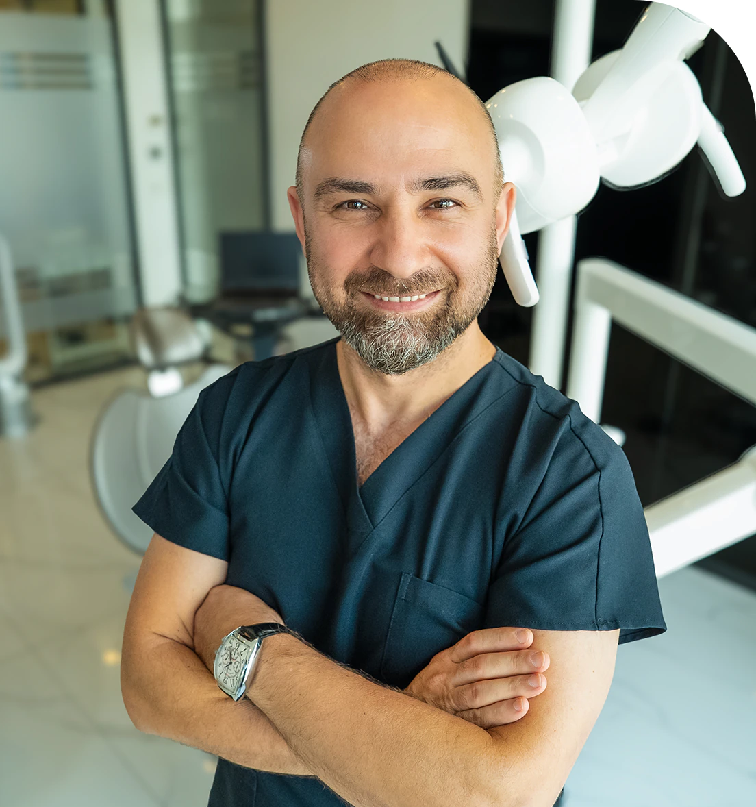
Dr. Dt. Ömer Atar provides care at his private clinic in Istanbul, combining his academic background and many years of clinical experience in dentistry. With a treatment approach that integrates aesthetic and functional solutions, he aims to achieve natural, balanced, and long-lasting results through personalized planning for each patient. Dr. Atar completed his education at the Marmara University Faculty of Dentistry, which he began in 1994, graduating in 2000. He continued his academic work in the Department of Prosthodontics at the same university. In 2007, he earned his PhD degree with his scientific thesis on implants and implant-supported prostheses in patients undergoing distraction osteogenesis. His academic expertise deepened in the fields of metal-free aesthetic systems, implant-supported prostheses, and cosmetic dentistry. Integrating modern technologies and digital dentistry into his practice, Dr. Ömer Atar achieves high success rates in treatments such as smile design, All-on-4/All-on-6 implant treatments, laminate veneers, zirconium crowns, teeth whitening, and gingival aesthetics (gummy smile). As a member of EDAD (Turkish Academy of Aesthetic Dentistry), he embraces a holistic approach focused on modern, functional, and natural outcomes in aesthetic dentistry.


