
 Türkçe
 Français
 Deutsch

Canines, medically known as cuspids, are located at the four corners of the oral anatomy, have the longest root structure, and are the most mechanically resilient teeth of the chewing system. Commonly referred to as “eye teeth” among the public, these strategic structures not only function to tear food but also serve as guides that protect the posterior teeth from excessive load during jaw movements, and shape facial aesthetics by supporting the corners of the lips. Due to delays in their eruption timing, canines are the second most commonly impacted teeth after wisdom teeth, and they are vital organs whose retention in the mouth is crucial for maintaining functional integrity and whose loss is the most difficult to compensate.
Why Are Canines the Strongest Anchors in the Mouth?
When you look in the mirror and smile, the prominent teeth standing at the arch of your dental alignment are merely the visible part of the iceberg. Canines have the longest root structure compared to all other teeth in the mouth. If we take a lower jaw canine as an example, its total length can reach approximately 27 millimeters, and a significant portion of it—around 16 millimeters—is embedded within the jawbone. This tremendous root length provides the tooth with an incredible anchoring power within the bone.
This robustness plays a vital role in counteracting horizontal forces that arise during chewing or in cases like teeth grinding. When you slide your jaw to the right or left, the first tooth that makes contact and bears the load is typically the canine. We call this “canine guidance.” If these teeth are not in place and stable, all destructive forces are transmitted to the posterior molars or anterior incisors. Over time, this may lead to cracks, fractures, or gum recession in other teeth. So, your canines are not just ordinary teeth; they are cornerstones of your oral health.
Why Do Impacted Canines Fail to Erupt and Cause Problems?
In dentistry, after wisdom teeth, canines are unfortunately the most commonly impacted teeth. Our patients often ask why these teeth fail to erupt while all others have already emerged. The underlying reasons are usually complex, but we can list the most prominent factors.
Common causes for canines remaining impacted or failing to find their path include:
- Lack of space in the jaw
- Retention of the baby tooth
- Genetic factors
- Jaw cysts
- Root damage due to trauma
- Closure of the eruption pathway
- Early loss of baby teeth
Among the items in this list, the persistent presence (retention) of the baby tooth is the most frequently encountered problem. If the baby canine does not loosen and fall out on time, the permanent tooth underneath must seek a new path. This search may sometimes lead it deep into the palate or towards the roots of neighboring teeth. Especially around the ages of 9–10, closely monitoring this process can prevent significant future complications.
How Does the Diagnosis Process Progress with Tomography?
When we suspect the presence of an impacted tooth, the simple panoramic X-rays used in the past would provide limited information. In two-dimensional images, we could only see whether the tooth was high or low, but it was difficult to understand its depth—whether it was closer to the lip or the palate. This could turn surgical planning into a guessing game.
Today, however, Cone Beam Computed Tomography (CBCT), one of the greatest blessings of modern technology, comes into play. Thanks to this technology, we can generate a three-dimensional map of the impacted tooth. We can see the tooth’s angle, whether it is damaging neighboring roots, with millimetric precision.
Critical information provided by tomography includes:
- Exact location of the tooth
- Inclination of the root
- Relationship with adjacent teeth
- Bone density
- Presence of cysts
- Status of surrounding tissues
When canines remain impacted, they carry the risk of resorbing the roots of adjacent incisors. With tomography, we can detect this risk early and take preventive measures before losing neighboring teeth. While determining the treatment strategy, we find the answer to the question, “Is it worth saving this tooth, or is extraction the healthier option?” in these 3D images.
What Are the Treatment Options for Impacted Teeth?
Once we confirm that a canine is impacted, we chart a course depending on the patient’s age, tooth position, and expectations. Our priority is always to preserve the natural tooth and bring it to its proper place. However, each case is unique, and sometimes we must follow different paths.
Basic treatment approaches applied in clinical practice include:
- Monitoring only
- Surgically uncovering the tooth and guiding it with braces
- Performing autotransplantation
- Extracting the tooth and placing an implant
- Extracting the tooth and closing the space
Among these options, the most frequently preferred and ideal method is to uncover the tooth and guide it into place with orthodontic forces. However, if the tooth is very deep, the patient is older, or there are root abnormalities, other options are considered. When making a decision, it’s essential not to rush and to discuss the pros and cons in detail.
How Is the Tooth Rescued with Orthodontic Treatment?
If we determine from tomography that the tooth is in a recoverable position, we initiate a procedure called “forced eruption.” This process requires an impressive collaboration between surgery and orthodontics. First, under local anesthesia, a small window is opened, and the bone and gum tissue over the impacted tooth are lifted. At this stage, a small button (bracket) with a chain or wire attached is bonded to the tooth.
Then, with the support of braces, the impacted tooth is gradually pulled toward its proper position, millimeter by millimeter. This process requires patience. While the tooth moves within the bone, it must remain vital, and the surrounding gum tissue must also adapt healthily. Since rapid movement can damage the tooth, forces are applied very slowly and in a controlled manner. Although treatment duration varies depending on the case’s difficulty, the reward of regaining the patient’s own vital tooth makes all the effort worthwhile.
Is Transplanting Your Own Tooth (Autotransplantation) Possible?
Sometimes, the impacted tooth is in such a reversed, distant, or horizontal position that pulling it into place with braces is impossible or highly risky. At this point, one of the most fascinating areas of medicine, “autotransplantation,” comes into play. This procedure is essentially an organ transplant; the difference is that both the donor and recipient are the patient.
In this procedure, the impacted tooth is surgically removed from its location as a single piece without damaging its root. Simultaneously, an artificial socket is prepared in the area where the tooth should be, and the tooth is transferred into this new socket.
Conditions required for successful autotransplantation include:
- Sufficient bone volume
- Appropriate root structure
- Absence of infection
- Delicate surgical technique
- Good healing potential
There are always risks of the tooth not fusing with the bone or root resorption after transplantation. However, when successful, we use the patient’s own living tissue instead of artificial materials like implants. These teeth can function in the mouth for years, even a lifetime.
When Is Root-End Surgery Necessary?
Canines may become infected due to deep cavities or trauma and may require root canal treatment. Root canal therapy is usually sufficient to save the tooth. However, in some persistent cases, the infection at the root tip does not heal or turns into a cyst. Sometimes, the root canals are so calcified that it’s impossible to reach the root tip with instruments.
In such cases, we resort to root-end surgery, called “apical resection.” In this procedure, a small incision is made in the gum to access the root tip directly.
Cases where apical resection is necessary include:
- Cyst formation at the root tip
- Failed root canal treatment
- Fracture at the root tip
- Excessively curved root structure
- Broken instruments inside the canal
- Overfilled canal fillings
During the procedure, infected tissue is cleaned, and a few millimeters of the root tip are removed. The canal opening at the root tip is then sealed with bioceramic filling materials that are highly compatible with the body. These materials prevent bacterial leakage and accelerate bone healing. Since canine roots are very long, working in this area requires great precision; attention must be paid to nearby nerves and anatomical spaces.
How Are Aesthetic Problems Solved in Smile Design?
Canines are not only functional but also one of the most critical elements in determining facial character aesthetically. Very pointed canines can give a harsher expression, while worn and flattened tips may make one look older. Sometimes, canines may be slightly rotated, have a darker color, or display asymmetric gums.
If the patient’s main concern is aesthetics and they do not prefer long-term orthodontic treatments, porcelain laminates (veneers) offer a perfect solution.
Situations where laminates are advantageous include:
- Discolorations
- Mild crowding
- Gaps between teeth
- Shape anomalies
- Surface wear
- Fractures or cracks
With these thin porcelain veneers, it is possible to change the tooth’s color, form, and size in just a few sessions. Especially for slight rotations in canines, it is a highly effective method to mask the irregularity with minimal abrasion and align the tooth into the ideal smile curve. However, it is crucial to preserve the natural structure of the tooth. We always avoid removing excessive healthy tooth tissue solely for aesthetic reasons.
Why Is Early Diagnosis So Important?
All the complex surgeries, transplants, and prolonged treatments we described above demonstrate the power of modern dentistry to solve problems. However, the best treatment is the one that resolves the issue before it escalates. Most canine-related problems can be managed much more simply when detected early.
Especially when children reach the ages of 9–10, a check-up before the permanent canines erupt is of vital importance. A simple X-ray taken at this age shows whether the tooth is progressing normally. If there is deviation or the baby tooth is resisting shedding, a timely, simple extraction or space maintainer application can save the child from major surgeries in the future.
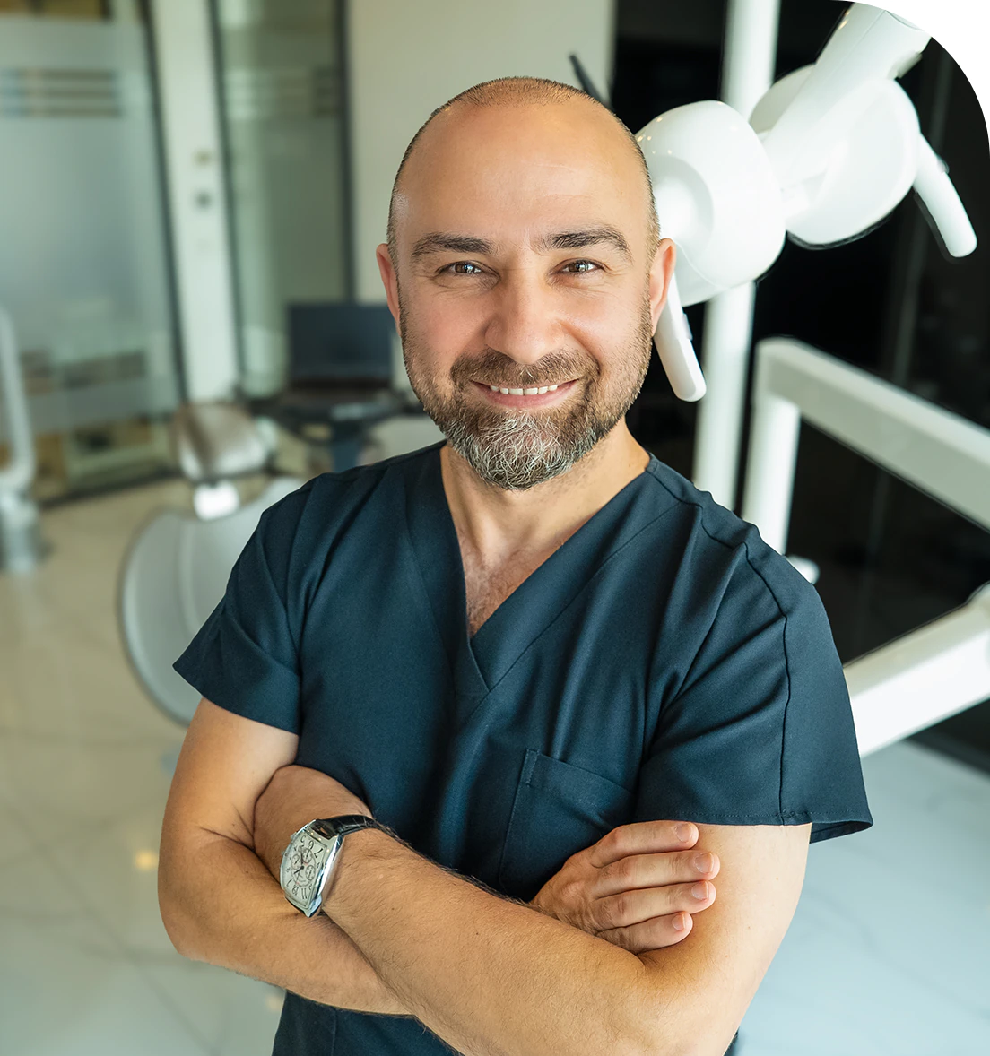
Dr. Dt. Ömer Atar provides care at his private clinic in Istanbul, combining his academic background and many years of clinical experience in dentistry. With a treatment approach that integrates aesthetic and functional solutions, he aims to achieve natural, balanced, and long-lasting results through personalized planning for each patient. Dr. Atar completed his education at the Marmara University Faculty of Dentistry, which he began in 1994, graduating in 2000. He continued his academic work in the Department of Prosthodontics at the same university. In 2007, he earned his PhD degree with his scientific thesis on implants and implant-supported prostheses in patients undergoing distraction osteogenesis. His academic expertise deepened in the fields of metal-free aesthetic systems, implant-supported prostheses, and cosmetic dentistry. Integrating modern technologies and digital dentistry into his practice, Dr. Ömer Atar achieves high success rates in treatments such as smile design, All-on-4/All-on-6 implant treatments, laminate veneers, zirconium crowns, teeth whitening, and gingival aesthetics (gummy smile). As a member of EDAD (Turkish Academy of Aesthetic Dentistry), he embraces a holistic approach focused on modern, functional, and natural outcomes in aesthetic dentistry.


