
 Türkçe
 Français
 Deutsch

Tooth anatomy refers to the structural features and functional parts of a tooth. Teeth are made up of different layers including enamel, dentin, pulp, cementum, and periodontal tissues. Each structure plays a role in maintaining functions such as chewing, speaking, and aesthetics.
Enamel is the outermost layer of the tooth and the hardest tissue in the body. The underlying dentin has a softer structure and is closer to the nerve endings. The pulp is located at the center of the tooth, contains vessels and nerves, and provides vitality and nourishment.
Cementum is the protective layer surrounding the tooth root and works together with the periodontal ligaments that connect the tooth to the jawbone. These connective tissues ensure the stability of the tooth and help in balancing chewing forces.
Teeth are classified by shape and function as incisors, canines, premolars, and molars. The front teeth serve a cutting function, while the back teeth grind. Proper understanding of tooth anatomy forms the basis for preventive and therapeutic dental practices.
| Things to Know | Information |
| Definition | Tooth anatomy includes the structural components of a tooth and their functions. Teeth are involved in the first step of digestion and serve functions like chewing, speaking, and aesthetics. |
| Main Parts | Crown: The visible part of the tooth in the mouth. Neck (cervical region): Located at the gum line. Root (radix): The part embedded in the jawbone. |
| Tissue Layers | Enamel: Covers the outer surface of the crown; the hardest structure in the body. Dentin: Located beneath enamel; yellowish and softer. Pulp: The living part containing nerves and vessels. Cementum: Covers the root surface. |
| Pulp Chamber | The hollow in the center of the tooth. Contains nerves, blood vessels, and connective tissue. The pulp extends both in the crown and root canals. |
| Root Canals | Thin canals that extend from the pulp chamber to the root tip. Require root canal treatment when infected. |
| Types of Teeth | Incisors: Front, cutting function. Canines: Pointed, for tearing. Premolars: Function both to cut and grind. Molars: Located in the back, used for grinding. |
| Number of Teeth | Adults have 32 permanent teeth: 8 incisors, 4 canines, 8 premolars, and 12 molars (including wisdom teeth). |
| Supporting Structures | Periodontal ligament: Fibers connecting the tooth to the jawbone. Alveolar bone: The jawbone surrounding the root. Gingiva: Soft tissue around the tooth. |
| Functions | Chewing and grinding, supporting speech, preserving facial aesthetics, contributing to the digestive system. |
| Oral Location | Teeth are symmetrically arranged in the upper (maxilla) and lower (mandible) jaws. Each jaw contains 16 permanent teeth. |
What Is Tooth Anatomy?
Tooth anatomy is the branch of science concerned with the basic layers and sections that make up teeth. Each tooth consists of three main parts:
- Enamel: The outermost layer of the tooth; hard and protective.
- Dentin: The softer layer beneath the enamel, closer to nerve endings.
- Pulp: Located at the center of the tooth; contains blood vessels and nerves.
A tooth is also divided into three main anatomical regions: the crown (visible part), neck, and root. The health of these structures is critical for overall oral health.
Why Is the Enamel Tissue, the Tooth’s Outer Shield, So Important?
When you look in the mirror, the shiny outer surface of your tooth—enamel—is the hardest substance your body produces. It’s harder than bone and is a mineral reservoir dense enough to be cut by diamond. However, this hardness doesn’t mean it’s unbreakable; rather, it’s brittle like glass. Under the microscope, enamel is composed of millions of small crystals arranged with incredible order into structures called “enamel prisms.” These prisms are aligned at specific angles from inside to outside.
This is the secret to clinical success. When a dentist removes decay for a filling, the remaining enamel must be shaped according to the prism orientation. If the supporting tissue is lost or the margin contradicts the prism direction, the area may fracture under chewing forces. That simple-looking margin polishing is actually a requirement of this microscopic engineering.
Enamel also contains weaker developmental regions called “enamel tufts.” These areas, like weak mortar in a wall, are more vulnerable. Bacteria and acids exploit these paths to infiltrate the tooth. The acids and adhesives we use in restorations create microscopic locks by penetrating this prismatic structure. That’s why keeping the tooth dry during procedures is vital; this delicate locking system is instantly affected by moisture.
Main factors damaging enamel:
- Acidic beverages
- Aggressive brushing
- Teeth grinding
- Nail biting
- Chewing ice
- Reflux disease
How Does the Dentin Layer, the Source of Tooth Sensitivity, Work?
Right beneath the hard protective enamel lies dentin—the main body of the tooth—yellowish, alive, dynamic, and sensitive. Its mineral content is lower (~70%), making it more flexible and acting as a shock absorber during chewing. But the most fascinating and troublesome feature of dentin is its tubular structure.
Think of dentin as millions of microscopic straws, each containing fluid and directly connected to the tooth’s nerve (pulp). When you drink cold water or eat something sour, the fluid shifts, causing sharp pain. If enamel wears off or the gum recedes, these tubules are exposed to the environment, and every thermal change triggers a signal to the nerve.
Dentin also ages. In youth, tubules are wide, so decay progresses quickly, and sensitivity is high. As we age or experience micro-traumas, new inner dentin forms—“secondary dentin”—narrowing the pulp and blocking tubules. This is a natural defense, reducing sensitivity but complicating root canal treatment.
Triggers of dentin sensitivity:
- Cold air
- Hot tea
- Citrus juices
- Sweets
- Hard brushing
- Whitening products
Why Is the Cementum Covering the Tooth Root So Delicate?
Only one-third of the tooth is visible; the root, two-thirds, is embedded in the bone. The root isn’t covered by enamel but by a thin, bone-like tissue called cementum, which provides an anchoring surface for fibers that connect the tooth to bone.
Cementum is not as hard as enamel. In gum recession, root surfaces become exposed and defenseless. Improper horizontal brushing quickly erodes cementum. When it’s worn away, the underlying dentin and nerve tubules are fully exposed, causing chronic sensitivity and rapid root decay.
During tartar cleaning or deep gum treatment (curettage), dentists aim to preserve cementum while removing infected tissue. Loss of this layer impairs healing and reduces the tooth’s biological integration.
Signs of damaged cementum:
- Yellowing on the root
- Tenderness to touch
- Gum recession
- Pain during brushing
- Extreme cold sensitivity
Why Is Mapping the Root Canal System Vital in Endodontic Therapy?
When you visit the clinic with severe tooth pain and need a root canal, the procedure involves cleaning a complex cave-like system inside the tooth. Most patients imagine the root as a straight pipe, but real anatomy is far more intricate. Canals split, merge, and may exit through multiple tiny pores (apical deltas).
Variations, especially in molars and lower front teeth, determine treatment success. A single canal might hide a second (Types 4 & 5), or a canal may bifurcate mid-root and merge again (Types 6 & 7). Here, CBCT (Cone Beam CT) comes in, offering 3D imaging beyond traditional X-rays.
Dental microscopes and magnifiers help identify hidden canals or “isthmuses”—narrow connections between canals. Bacteria love these passages. If not cleaned, pain or abscess may persist or recur years later.
Common root canal variations:
- Double canals
- C-shaped canals
- Extra roots
- Lateral canals
- Apical deltas
- Fused canals
How Does Periodontium Support Prevent Tooth Loosening?
Teeth are not nailed into the bone. If directly fused, each bite would transmit shock to the skull. Instead, teeth hang like hammocks in a **periodontium** support system. Its key component is the periodontal ligament (PDL)—thousands of tiny fibers.
One end of each fiber connects to cementum, the other to bone. They allow microscopic movement and absorb forces. These fibers also provide tension that keeps the bone alive.
When gum disease begins, tartar and bacteria invade the gum pocket. The body’s defense dissolves its own tissues. Fibers break, then bone resorbs. Patients may feel no pain but see bleeding. Eventually, even healthy teeth loosen and fall. Thus, tooth anatomy includes this support system as well.
Factors harming periodontium:
- Tartar buildup
- Poor oral hygiene
- Smoking
- Diabetes
- Genetics
- Stress
How Does Malocclusion Strain the Jaw Joint?
Tooth anatomy isn’t just about single units; it also includes how they contact their counterparts in the opposite jaw—this is **occlusion**. Ideally, one tooth touches two opposing ones for balanced force distribution.
But even a slightly high filling or a missing tooth can disrupt this harmony. If one tooth hits prematurely, it receives excessive force—causing pain or cracks, and more importantly, affecting the TMJ (temporomandibular joint).
The joint, located in front of your ears, may develop disk displacement, clicking, or locking. Some patients seek ENT or neurology care for headaches or ear pain, unaware the root cause is malocclusion. Therefore, all prosthetic or restorative work must follow anatomical rules.
Symptoms of occlusal imbalance:
- Jaw joint pain
- Morning fatigue
- Unexplained headaches
- Tooth wear
- Gum recession
- Neck/back pain
How Is Baby Tooth Anatomy Different from Permanent Teeth?
A common parental mistake is to neglect **baby teeth**, assuming they’ll fall out. However, their anatomy makes them more sensitive and risky. Enamel and dentin are much thinner, while the pulp chamber is proportionally larger.
Clinically, this means a small cavity can quickly reach the nerve. While it might take months in adults, it may abscess in weeks in a baby tooth. In primary molars, nerves lie close to the surface—especially on the sides—making treatment risky.
Baby tooth roots dissolve over time to make way for permanent teeth. But they also act as space maintainers. Early loss allows neighboring teeth to shift, blocking the eruption path of the permanent tooth, leading to orthodontic problems. X-rays must monitor root resorption and eruption timing.
Differences of baby teeth:
- Whiter color
- Thinner enamel
- Larger pulp chamber
- Finer roots
- Faster decay
- Root resorption
- Clinical Perspective
The main takeaway from this comprehensive analysis is: dentistry is not just skill but an artistic mastery of anatomy. Diagnosis is half the treatment. Spotting a Type 6 canal, locating a child’s pulp exposure, or identifying jaw pain from a high filling requires this knowledge.
Modern tools (CBCT, microscopes) help us visualize anatomy, but true preventive care begins with your awareness. Knowing how acids erode enamel or gum recession exposes vulnerable cementum may transform your oral hygiene. Preserving this anatomical perfection is always easier and more valuable than repairing it. Demanding treatments that respect biomechanical rules and natural structure is the best step toward long-term oral health.
Frequently Asked Questions
What layers make up tooth anatomy?
Tooth anatomy has three main layers: enamel, dentin, and pulp. Enamel is the outer hard layer, dentin is in the middle, and pulp contains vessels and nerves in the center.
What is the function of enamel in tooth anatomy?
Enamel is the tooth’s outer layer and the hardest tissue in the body. Its main role is to protect against chewing forces and acid erosion, but it can wear down over time.
How is dentin related to sensitivity?
Dentin lies under the enamel and contains tiny tubules. When enamel wears away, these become exposed and sensitive to heat, cold, and other stimuli.
What happens when the pulp layer becomes inflamed?
Since the pulp contains nerves and blood vessels, inflammation results in a painful condition called pulpitis. If untreated, it may lead to tooth loss.
How does tooth anatomy change with age?
Enamel thins, dentin thickens, and the pulp chamber narrows with age. This can reduce sensitivity but increase decay risk.
Does tooth anatomy differ between men and women?
While overall structure is the same, studies show men tend to have larger, stronger teeth, while women may be more prone to gum recession.
How does tooth anatomy affect orthodontic treatment?
Root length, bone density, and pulp status directly affect treatment duration and success. Healthy anatomy responds better to orthodontics.
Which part is most affected by decay?
Cavities usually start by eroding enamel and then quickly progress to dentin, which is softer and more porous.
Why is tooth anatomy important in cosmetic dentistry?
Aesthetic treatments consider enamel color, tooth shape, and symmetry. Materials are chosen to match enamel structure for a natural look.
How do genetics affect tooth anatomy?
Genes influence tooth size, shape, enamel thickness, and alignment. Some people may have congenital enamel defects or missing teeth.
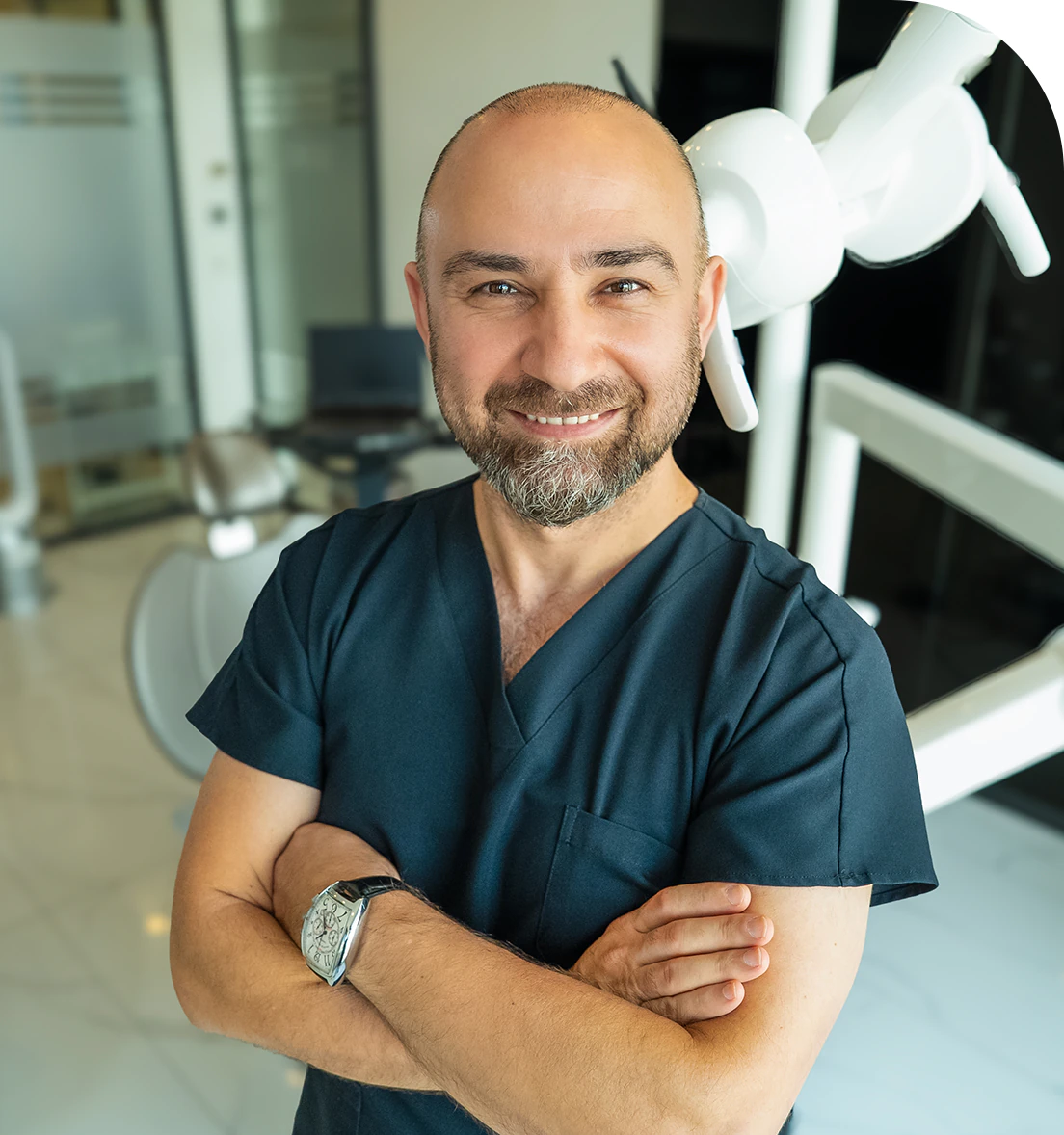
Dr. Dt. Ömer Atar provides care at his private clinic in Istanbul, combining his academic background and many years of clinical experience in dentistry. With a treatment approach that integrates aesthetic and functional solutions, he aims to achieve natural, balanced, and long-lasting results through personalized planning for each patient. Dr. Atar completed his education at the Marmara University Faculty of Dentistry, which he began in 1994, graduating in 2000. He continued his academic work in the Department of Prosthodontics at the same university. In 2007, he earned his PhD degree with his scientific thesis on implants and implant-supported prostheses in patients undergoing distraction osteogenesis. His academic expertise deepened in the fields of metal-free aesthetic systems, implant-supported prostheses, and cosmetic dentistry. Integrating modern technologies and digital dentistry into his practice, Dr. Ömer Atar achieves high success rates in treatments such as smile design, All-on-4/All-on-6 implant treatments, laminate veneers, zirconium crowns, teeth whitening, and gingival aesthetics (gummy smile). As a member of EDAD (Turkish Academy of Aesthetic Dentistry), he embraces a holistic approach focused on modern, functional, and natural outcomes in aesthetic dentistry.


