
 Türkçe
 Français
 Deutsch
 Nederlands
 Svenska

Premolars are strategically positioned elements of the chewing system, located between the canines and molars in the oral anatomy, facilitating the transition from the tearing to the grinding phase of food processing. While maintaining the aesthetic continuity of the anterior region, these teeth also absorb the high chewing pressures of the posterior region. They play a crucial role in preserving functional occlusion and the balance of the temporomandibular joint. Due to the high biomechanical stress they endure and their unique anatomical structure, premolars are prone to cracks. Therefore, advanced restorative methods such as inlays, onlays, and overlays—which preserve tooth structure and enhance durability—are the most effective clinical solutions for ensuring the long-term function of these teeth.
What role do premolars play in the mouth?
If you think of your mouth as a construction site, each group of teeth can be compared to a specific type of machine. The incisors act like scissors, cutting food. The canines grip and tear like powerful hooks. The molars at the back function like giant millstones that crush food. So what do the premolars, positioned in the middle of this system, do? They serve as “transfer gears.”
Premolars manage the transition from the food-breaking phase to the grinding phase. Located in the most dynamic and mobile region of the jaw, the load they endure is far greater than one might assume. They handle vertical crushing forces as well as horizontal forces that occur when the jaw moves side to side. Due to this strategic position, they are considered the most critical turning point of the chewing system. If there’s a problem at this turn, the entire chewing mechanism may be disrupted.
What is the significance of the grooves and cusps (occlusal morphology) on these teeth?
When you run your tongue over a premolar, you’ll notice it doesn’t feel like a flat surface but rather like a terrain of valleys and peaks. This is called “occlusal morphology,” and it’s no coincidence. It represents one of nature’s most efficient engineering designs. The cusps (elevations) and fossae (depressions) on the tooth surface function in a key-and-lock harmony with the opposing teeth in the other jaw.
The primary purpose of this design is to control the enormous forces generated during chewing. Think of a nutcracker: if you apply force directly to the center, the shell cracks easily; if the pressure is applied unevenly from the sides, the nut may slip out. The specialized shapes on premolars capture the force and transmit it directly along the tooth’s “long axis”—the strongest direction—toward the root. This enables the tooth to function like a firmly anchored nail, without shifting sideways. If this natural form is disrupted, forces may disperse incorrectly, compromising the tooth’s stability.
Why does Cracked Tooth Syndrome affect premolars more frequently?
One of the most common yet hardest-to-diagnose complaints we encounter in clinical practice is “Cracked Tooth Syndrome.” Unfortunately, premolars are its most frequent target—due to basic physics and anatomy. Compared to molars, premolars are more delicate and slender in structure. Yet the load they bear is nearly equal to that of molars.
Premolars have a deep natural groove between their two cusps. When biting into a hard object, this groove can act like a wedge, forcing the two sides of the tooth apart. If there is a large existing filling or if the patient suffers from bruxism (teeth grinding), the tooth cannot withstand this flexing motion. Over time, hairline fractures—thinner than a strand of hair—may develop. This is similar to splitting a log with an axe; with every bite, the crack deepens and advances toward the pulp, the living tissue of the tooth.
What signals does the body give when a premolar cracks?
A cracked tooth can be confusing for both patient and clinician, as the pain is not always constant. With decay, a tooth typically throbs persistently. But a cracked tooth behaves differently—its pain comes and goes, like playing hide-and-seek. Patients often say, “Doctor, I can’t point to exactly where it hurts, but I know something is wrong.”
The most common symptoms of this syndrome are:
- Sudden, sharp pain during chewing
- Relief when pressure is released
- Sudden sensitivity to cold drinks
- Discomfort from sugary foods
- Inability to pinpoint the pain’s origin
- Pain that occurs only when biting at a specific angle
Why is diagnosis so difficult and what methods do dentists use?
Detecting a crack in a tooth can be like finding a needle in a haystack. X-rays are great at showing bone and hard tissues, but they often fail to reveal extremely thin, incomplete cracks. These cracks may be hidden under fillings or start below the gum line, making visual inspection nearly impossible.
At this point, we dentists must act like detectives. After listening to the patient’s symptoms, we use a special tool called a “bite stick.” With this device, we apply pressure to each cusp of the suspect tooth individually. The goal is to locate the crack and reproduce the characteristic pain. If the patient reacts not when biting down, but when releasing the pressure and says, “That’s it!”, we consider it the strongest evidence of a cracked tooth. The crack opens when pressure is applied and snaps shut when released, pinching the nerve and causing sharp pain.
Why are inlays, onlays, or overlays preferred over traditional fillings?
Traditional fillings work well for small to moderate cavities. However, when dealing with severely damaged or crack-prone premolars, classic fillings can sometimes worsen the situation. White (composite) fillings cured inside the mouth tend to shrink under light. This shrinkage pulls the opposing tooth walls together, creating constant tension. Already weakened walls may eventually crack under this stress.
This is where “indirect restorations” such as inlays, onlays, and overlays come into play. These restorations are fabricated outside the mouth in a lab from porcelain or reinforced materials. They act like durable puzzle pieces that restore the missing portion of the tooth. Their main advantage is that they protect the tooth by wrapping around its weak areas from the outside, rather than merely filling the inside. Onlays and overlays, in particular, cover the cusps like a protective cap. This “cusp coverage” effect physically prevents the tooth from flexing and the crack from propagating. Thus, these restorations are not just fillings—they are protective shields that reinforce the tooth’s biomechanical structure.
What are the differences between inlays, onlays, and overlays?
These terms can confuse patients, but they are simply technical classifications based on how much of the tooth is being covered. All are fabricated in the lab with the same precision and technology, but the coverage varies. Your dentist will determine the appropriate one based on the extent of the damage.
These types include:
- Inlay
- Onlay
- Overlay
An inlay is used when the cusps are intact and decay is limited to the central grooves—making it most similar to a filling. An onlay is chosen when the damage involves one or more cusps, covering and protecting them. An overlay is the most extensive type, used when the entire chewing surface is compromised and needs to be completely covered.
What risks are associated with untreated premolars?
Premolar problems that are ignored or masked with painkillers under the assumption that “it’ll pass” often evolve into more serious and costly conditions over time. A small crack that starts in the enamel can deepen with each bite. Once it reaches the pulp, conservative treatment is no longer possible, and root canal therapy becomes necessary.
But the risks don’t stop there. Neglect can lead to:
- Vertical fractures extending to the root
- Loss of the tooth requiring extraction
- Infections and abscesses in the jawbone
- Facial swelling and severe pain
- The need for more expensive treatments like implants or bridges
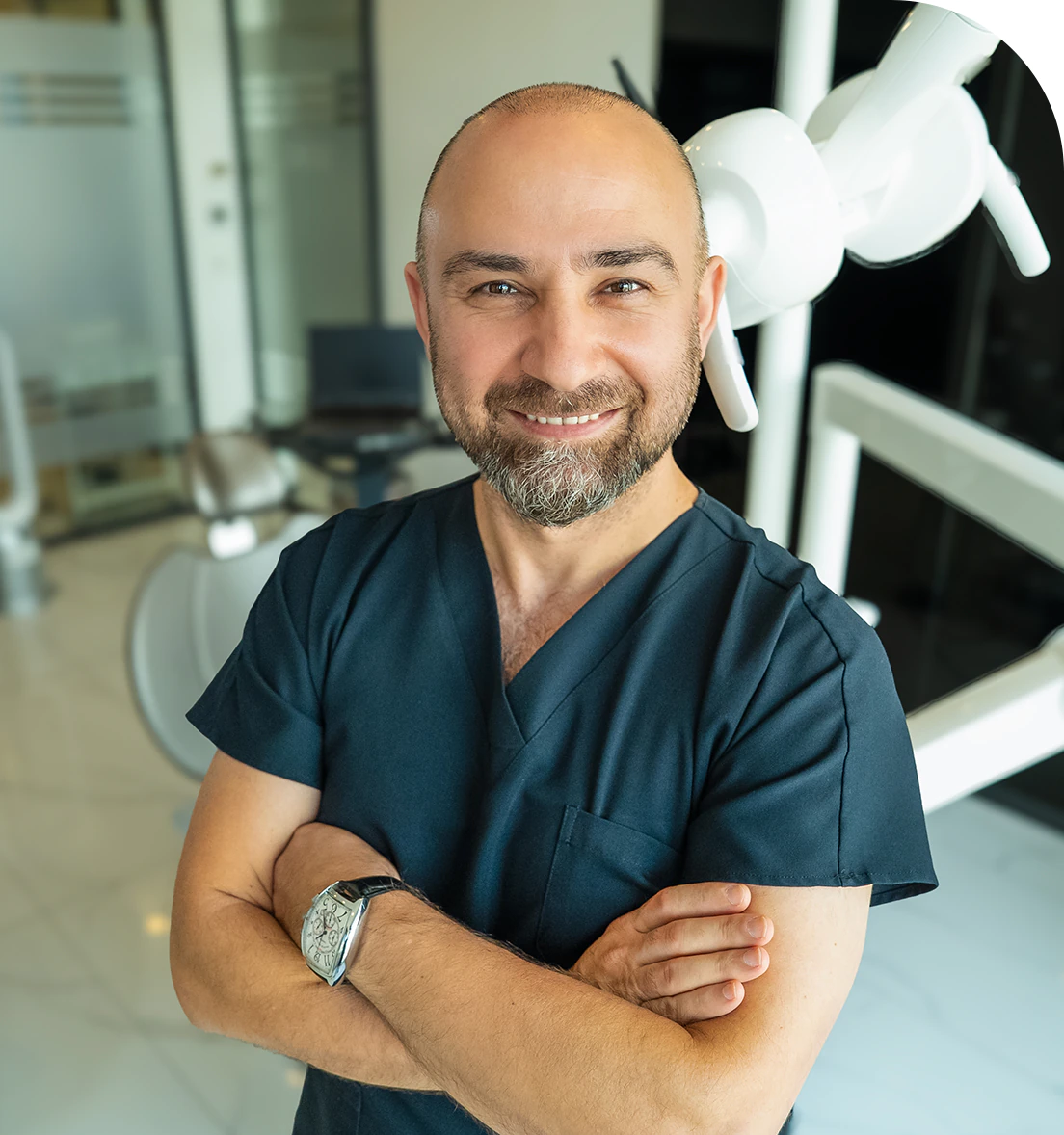
Dr. Dt. Ömer Atar provides care at his private clinic in Istanbul, combining his academic background and many years of clinical experience in dentistry. With a treatment approach that integrates aesthetic and functional solutions, he aims to achieve natural, balanced, and long-lasting results through personalized planning for each patient. Dr. Atar completed his education at the Marmara University Faculty of Dentistry, which he began in 1994, graduating in 2000. He continued his academic work in the Department of Prosthodontics at the same university. In 2007, he earned his PhD degree with his scientific thesis on implants and implant-supported prostheses in patients undergoing distraction osteogenesis. His academic expertise deepened in the fields of metal-free aesthetic systems, implant-supported prostheses, and cosmetic dentistry. Integrating modern technologies and digital dentistry into his practice, Dr. Ömer Atar achieves high success rates in treatments such as smile design, All-on-4/All-on-6 implant treatments, laminate veneers, zirconium crowns, teeth whitening, and gingival aesthetics (gummy smile). As a member of EDAD (Turkish Academy of Aesthetic Dentistry), he embraces a holistic approach focused on modern, functional, and natural outcomes in aesthetic dentistry.


