
 Türkçe
 Français
 Deutsch

Impacted wisdom tooth extraction is the surgical removal of wisdom teeth that have not fully erupted or have developed in an incorrect position within the jawbone. These teeth can often lead to problems such as pain, infection, cyst formation, and disruption of tooth alignment.
The extraction of impacted teeth is planned after a detailed evaluation using a panoramic X-ray. Depending on the position of the tooth, the procedure is performed under local anesthesia, and the surrounding bone tissue is opened to remove the tooth. After the surgical extraction, the area is closed with sutures.
Mild swelling, pain, and difficulty in chewing may occur for a few days following the operation. Painkillers and antibiotics prescribed by the doctor should be used; ice application, a soft food diet, and good oral hygiene are essential. Smoking adversely affects healing.
Impacted wisdom tooth extraction is typically completed in a single session; however, for multiple impacted teeth, intervention may be planned at intervals. The procedure is important to prevent more serious future jaw and dental health issues.
| What You Should Know | Information |
| Definition | Impacted wisdom tooth extraction is the surgical removal of third molars that are fully or partially embedded in the jawbone, often unerupted or not positioned correctly. Most commonly seen in the lower jaw. |
| Types of Impaction | – Fully impacted: The tooth remains completely within the bone. – Partially impacted: Part of the tooth has emerged through the gum. – Horizontal, vertical, angled impaction: Classified based on the eruption direction of the tooth. |
| Reasons for Extraction | – Gum infection (pericoronitis) – Pressure on adjacent teeth and risk of decay – Cyst formation – Jaw pain or swelling – Lack of space before orthodontic treatment – Recurrent abscess formation |
| Symptoms | – Pain during chewing – Swelling and redness in the gum – Limited mouth opening – Bad breath – Decay or sensitivity in adjacent teeth |
| Procedure | Performed under local anesthesia. The gum and, if necessary, bone over the tooth are removed. The tooth may be sectioned and extracted in parts. The procedure duration varies between 20–60 minutes depending on the position of the tooth. |
| Post-Surgery | – Swelling, mild pain, and bleeding within the first 24 hours – Cold compress should be applied – Antibiotics and painkillers are prescribed – Avoid smoking and hot foods – Do not rinse mouth on the first day |
| Healing Process | Takes approximately 7–10 days. Full bone healing may take several months. Pain and swelling during the first 3 days are normal. |
| Complications | – Alveolitis (dry socket): Occurs when the clot dislodges, causing severe pain – Temporary jaw locking – Temporary numbness in the lower lip (especially with lower impacted teeth) – Development of infection and inflammation |
| Care and Recommendations | – Do not spit or rinse the mouth within the first 24 hours – Do not use straws – Opt for soft and lukewarm foods – Protect the surgical site while brushing |
| Alternatives | If the impacted tooth does not present significant symptoms, regular monitoring may be advised; however, if there is infection, pressure, or cyst risk, extraction is mandatory. |
| Long-Term Follow-Up | Sutures are typically removed after 7 days. Follow-up checks ensure proper wound healing and monitor for infection. |
What Is Impacted Wisdom Tooth Extraction?
Impacted wisdom tooth extraction is the surgical removal of wisdom teeth that have not fully erupted or have emerged at an incorrect angle within the jawbone. These teeth may cause pain, infection, decay, or misalignment of the teeth. The procedure is done under local anesthesia and generally involves a minor surgical intervention to remove the tooth. Healing duration varies by individual.
Why Do Impacted Wisdom Teeth Become a Problem, and What Does “Impacted” Mean?
Wisdom teeth, also known as third molars, are the last teeth located at the back of the dental arch. Over human history, dietary habits have changed from raw and tough foods to softer and processed ones, leading to an evolutionary reduction in jaw size. However, the number of teeth has remained the same. This mismatch — having fewer spaces in a smaller jaw for the same number of teeth — results in wisdom teeth not finding space to emerge and becoming trapped within the bone.
The impaction of a tooth does not always mean it will cause pain, but it can be a sign of silently progressing problems. When a tooth cannot find a way to erupt, it may press against the adjacent tooth while trying to emerge, rotate horizontally, or even remain completely inverted. What we refer to as “fully impacted” teeth are completely under the bone and gum and are not visible from the outside. However, the more clinically problematic group is usually the “partially impacted” teeth. A part of these teeth has emerged into the oral cavity, but the rest remains under the gum. This small opening creates a perfect tunnel for bacteria and food debris to enter under the gum. In this blind spot that a toothbrush cannot reach, bacteria accumulate and eventually create a ground for serious infections.
What criteria are used to decide whether these teeth should be extracted?
Our patients sometimes ask, “I have no pain—should I still have it extracted?” This is a critical question that varies depending on the patient. In the modern approach to dentistry, if a tooth is completely embedded in the bone, has no pathological formations like cysts or tumors around it, is not damaging the roots of adjacent teeth, and causes no complaints in the patient, it does not necessarily need to be extracted. In such cases, we implement a “monitoring” protocol. However, monitoring does not mean “forget and move on”; regular radiological checks are required to track the position of the tooth.
However, in some situations, the decision to extract becomes inevitable. Especially in partially impacted teeth, frequent gum inflammation (pericoronitis), bad breath, and pain radiating to the ear and neck are considered “red flags.” Additionally, if the impacted tooth is leaning against the healthy second molar in front of it, it can cause root resorption or lead to decay. In such cases, we must sacrifice the impacted tooth to protect the healthy one.
The main reasons that lead us to decide on extraction include:
- Recurrent gum infections
- Caries risk in neighboring teeth
- Cyst formation
- Suspicion of tumors
- Need for orthodontic treatment
- Unexplained jaw pain
- Prosthetic planning needs
Why are preoperative X-rays and CT scans so important?
Before starting surgery, we want to know exactly what we will face down to the millimeter. Performing “blind surgery” goes against surgical principles. That’s why during the first examination, we always take a panoramic X-ray, which provides a wide view of the entire jaw. This film serves as a map, showing us the general position of the tooth, bone level, and its relationship with other teeth. For most standard extractions, this map is sufficient.
However, in some cases, a two-dimensional map can be misleading. Especially in the lower jaw, the roots of the impacted tooth may appear dangerously close to the main nerve canal (mandibular nerve) that provides sensation to the lip. From the panoramic film, we cannot determine whether the roots are above, behind, or going directly through the nerve. This is where 3D tomography (CBCT) becomes crucial. The scan shows us the tooth from every angle and layer by layer. We can clearly see the root curves (curvatures), the exact distance to the nerve, and the bone density.
This detailed examination completely changes our surgical strategy. If the root is in contact with the nerve, we plan to section the tooth and remove it piece by piece rather than as a whole. In this way, we can remove the tooth like pulling a hair from butter, without applying pressure to the nerve. So, X-rays and tomography are not just films—they are the insurance policy for a safe surgery.
How does the tooth’s position affect the difficulty of the surgery?
Not all impacted tooth surgeries are equally difficult. The different stories our patients hear from friends like “Mine took 5 minutes” or “Mine took an hour” stem entirely from the tooth’s position within the bone. There are scientific classifications we use to determine this difficulty.
The most important of these is the angle of the tooth. If the tooth is positioned vertically like the others, our job is relatively easier. But if the tooth is tilted forward, leaning backward, or in the most difficult case, lying completely horizontally, things get more complicated. A horizontally positioned tooth is like a subway car that has tunneled through the bone. You can’t just pull it straight up, because the bone above and the tooth in front prevent it. In these “complicated” cases, we must open a wider surgical field and section the tooth into pieces using surgical techniques.
Another criterion is how deep the tooth is. The more the tooth is embedded within the ascending branch (ramus) of the jawbone, the harder it is to reach. In the classification known as Pell and Gregory, “Class 3” teeth are those located completely within the bone and far back, representing the highest level of surgical difficulty, requiring skill and experience. Identifying these cases is crucial for estimating surgery time and preparing the patient for potential outcomes like swelling.
What are the anesthesia options and what will be felt during the procedure?
Sitting in the dentist’s chair is a source of stress for many people—we are aware of that. But thanks to modern anesthesia methods, pain is no longer a parameter to fear. The vast majority of impacted tooth surgeries are comfortably performed under local anesthesia, meaning only the affected area is numbed. Once the injection is done, all sensation in that area disappears.
Here, our patients should understand the difference between “feeling” and “pain.” Local anesthesia eliminates pain 100%, but it does not entirely eliminate sensations of touch and pressure. So during the procedure, you may feel your doctor pushing or applying pressure on the tooth, or notice vibrations—but these sensations will not hurt. Understanding this distinction helps manage stress while in the chair.
However, in some cases, local anesthesia alone may not be enough. If the patient has severe dental phobia, a strong gag reflex, or if four difficult impacted teeth need to be extracted in one session, we consider “Sedation” or “General Anesthesia” options for patient comfort. Sedation involves administering medication through an IV to bring the patient into a deeply relaxed, semi-sleep state. The patient can respond to commands but feels no fear and does not remember the procedure afterward. General anesthesia is a full sleep state and is typically performed in a hospital under operating room conditions.
Patient groups for whom we prefer sedation or general anesthesia include:
- Those with severe dentophobia (fear of dentists)
- Mentally disabled individuals
- Patients who cannot stay still due to physical disabilities
- Large cyst cases where local anesthesia is insufficient
- Situations where multiple complex extractions are planned in one session
- Patients with extremely limited mouth opening
- Individuals with an excessive gag reflex
Step-by-step, how is the surgical extraction procedure performed?
Impacted tooth surgery is a micro-surgical procedure performed with delicate techniques, not brute force. The goal is not only to extract the tooth but also to preserve the surrounding bone and gum tissue. The procedure typically begins with sterilization and anesthesia. Once the area is completely numb, we start the surgical process.
The first step is to gently separate the gum to access the tooth. This step, known as “raising a flap,” involves opening the gum like a flap over the bone to expose the surgical field. If the tooth is under the bone, we perform a procedure called “osteotomy” to remove bone. Using special tips and abundant sterile water cooling, we carefully remove the minimal amount of bone necessary to free the tooth. Cooling is critical because overheating the bone can impair healing.
The next step, especially in difficult teeth, is “odontotomy,” meaning the division of the tooth. Trying to extract a tooth that is horizontally positioned or has divergent roots in one piece can strain the jawbone. Instead, we divide the tooth into several pieces using special instruments. This way, each piece can be removed easily through the small window created, without causing trauma. This technique is based on the “divide and conquer” principle and both accelerates healing and protects the jawbone.
After the tooth and all its parts are removed, the remaining socket is meticulously cleaned. No cyst remnants, infected tissue, or bone debris should remain inside. A clean wound is the foundation of healthy healing. Finally, the opened gum is repositioned and closed with sutures. Stitches keep the wound stable and stop the bleeding.
Is immediate extraction performed if the gums are inflamed or there is swelling in the face?
There is a golden rule on this topic: “No surgical intervention is performed on a tooth with acute infection.” If the patient comes in with facial swelling, inability to open the mouth, fever, and throbbing pain (an acute abscess picture), our priority is not extraction but to control the infection. Because the infected area is acidic, local anesthetic agents cannot work effectively in this environment—meaning the tooth won’t go numb. Additionally, cutting into infected tissue may cause bacteria to enter the bloodstream and spread throughout the body.
In such cases, we first start antibiotic therapy. If the abscess is matured, we drain it with a small intervention to relieve the patient. We then send the patient home and ask them to use their antibiotics regularly. Once the infection subsides, the swelling decreases, and the condition becomes “chronic”—in other words, when the patient is comfortable—we schedule the extraction appointment. This waiting period is essential both to prevent pain and to ensure surgical success.
Antibiotic use is not limited to abscesses. Sometimes it is also used “prophylactically,” meaning for prevention. After difficult bone surgeries, we may prescribe a 5-day course of antibiotics post-operation for patients with low immunity to minimize the risk of infection.
What is “Dry Socket” and how can it be prevented?
Our patients’ biggest postoperative fear is “dry socket,” or medically, “alveolitis sicca.” Normally, when a tooth is extracted, the socket fills with blood and clots. This clot acts like a scab on a skin wound—it covers the bone underneath and initiates healing. In the case of a dry socket, however, this clot either never forms or is dislodged due to patient error.
Once the clot is gone, the underlying bone and nerve endings are exposed. This presents as severe pain radiating to the ear and a bad odor in the mouth 2–3 days after the extraction. Dry socket is not an infection but a healing disorder. Its treatment is done in the dental chair: the wound is cleaned, and special medicated pastes that instantly relieve pain are placed inside.
Factors that increase the risk of dry socket include:
- Smoking
- Drinking through a straw
- Frequent spitting
- Rinsing the mouth with water
- Hot food and drinks
- Use of birth control pills
- Touching the wound with the tongue
This is why we insistently say to our patients: “Do not spit or rinse during the first 24 hours.” Spitting creates a vacuum in the mouth that dislodges the precious clot.
Is swelling and bruising on the face normal after surgery?
Yes, especially after difficult impacted tooth surgeries involving bone removal, swelling (edema) on the face is expected and is a natural response of the body. The body sends fluids and cells to the surgical area to promote healing, which appears as swelling externally. Don’t worry—this is a sign that healing has started. Swelling typically peaks on the second day after surgery and starts to subside from the third day onward.
Sometimes, this swelling may be accompanied by “trismus,” or jaw locking. The patient finds it difficult to open their mouth. This is related to both the mechanical effect of the swelling and spasms in the chewing muscles. As the swelling goes down, mouth opening returns to normal.
To go through this process as mildly as possible, ice application is critical. On the day of surgery, applying ice externally on the cheek for 10–15 minutes with 10-minute breaks prevents excessive swelling. Also, sleeping with two pillows under the head in the first few days reduces edema by lowering blood pressure in the area. If the skin is sensitive, yellow-green bruising may be observed in the swollen area—this is due to blood leaking under the skin and resolves over time.
Is there a risk of nerve damage and will there be numbness in the lips?
The most sensitive complication of lower jaw wisdom tooth extractions is “paresthesia,” or temporary sensory loss. As mentioned above, if the roots of the tooth are very close to the lip nerve, swelling or manipulation during surgery can temporarily compress the nerve. This does not mean the nerve is severed. It just means nerve transmission is momentarily interrupted, like water flow in a hose being stepped on.
As a result, the patient may experience tingling or numbness in part of the lip or the tip of the chin. Most of these cases resolve spontaneously within days or weeks as the swelling subsides. Rarely, it may persist up to 6 months. During this period, we recommend B vitamin supplements that accelerate nerve repair. Permanent sensory loss is now very rare thanks to 3D imaging technologies and microsurgical methods. If your surgeon identifies a risky situation, they will certainly inform you before the operation.
What should be done and eaten to speed up the healing process?
If 50% of surgical success depends on the surgeon, the other 50% depends on the patient’s care. The care you apply at home directly affects your comfort. The first rule is bleeding control. You must bite firmly on the gauze your dentist placed for 20–30 minutes. Bite it to apply pressure and stop the bleeding. After removing the gauze, mild oozing may occur, which is normal; there is no need to place another gauze or rinse your mouth. Although the taste of blood is unpleasant, spitting increases bleeding—so swallowing is best.
Nutrition is also very important. Until the numbness wears off—approximately 2–3 hours—you should not eat anything, otherwise, you may bite your cheek or lip without realizing it. For the first 24 hours, avoid granular, hard, very hot, and spicy foods.
Recommended foods include:
- Warm soups
- Mashed potatoes
- Yogurt
- Pudding
- Ice cream
- Fruit juices
- Pasta
- Boiled vegetables
Foods to avoid include:
- Nuts
- Chips and crackers
- Bread with sesame seeds
- Spicy seasonings
- Carbonated drinks
- Boiling hot food
- Grainy rice dishes
As for hygiene, you can skip brushing your teeth on the first day, but from the next day onward, you should brush your other teeth normally. When approaching the extraction site, do so gently without touching it. You can start using the mouthwashes prescribed by your dentist or warm salt water 24 hours after surgery.
Using a syringe (mak) is an excellent method for cleaning the wound area from food debris. And most importantly, do not stop taking the prescribed medication just because “there is no pain”—continue taking it regularly until the pack is finished. Remember, good patient-doctor cooperation ensures that even the most difficult surgeries can be overcome smoothly.
Frequently Asked Questions
In which cases does the extraction of impacted wisdom teeth become mandatory?
If the tooth remains fully embedded in the jawbone and exerts pressure, pushes surrounding teeth, or poses a risk of infection, surgical extraction becomes mandatory. This is done to prevent cyst formation and jaw pain.
What risks may arise if impacted wisdom teeth are close to nerve tissue?
In teeth close to the lower jaw nerve, temporary or rarely permanent numbness may occur during extraction. Therefore, detailed panoramic or 3D imaging is important before the procedure.
Why does difficulty in opening the jaw develop after impacted tooth extraction?
Due to surgical trauma and edema in the surrounding tissues, the chewing muscles may enter temporary spasm. This condition may last for several days and can be alleviated with cold application.
How long should soft food be consumed after impacted wisdom tooth extraction?
For the first 48–72 hours, hot and hard foods should be avoided, and soft, warm foods should be preferred. This supports clot protection and wound healing.
How does smoking affect healing after impacted tooth extraction?
Smoking causes narrowing of blood vessels, reducing blood flow and delaying healing. It also significantly increases the risk of infection and “dry socket” (alveolitis).
What are the symptoms when alveolitis develops after wisdom tooth extraction?
Severe throbbing pain that starts 2–4 days after extraction, bad breath, and loss of the clot in the empty socket are the main symptoms of alveolitis. If left untreated, the duration of pain extends.
Why does infection risk increase if the top of the impacted tooth is partially visible?
In partially impacted teeth, food accumulation between the tooth and gum is easy. This area cannot be properly cleaned, leading to bacterial infection (pericoronitis).
Which methods are effective in preventing swelling after extraction?
Applying cold compresses externally at intervals during the first 24 hours reduces the risk of swelling. Keeping the head elevated and avoiding excessive mouth rinsing is also beneficial.
Is stitching necessary after the extraction of impacted wisdom teeth?
Stitches are placed in most surgical extractions. This is important for controlling bleeding, holding tissues together, and accelerating healing. Non-dissolvable stitches are usually removed after 7 days.
How long does the full healing process take after impacted tooth extraction?
Soft tissue healing is usually completed within 1–2 weeks. However, full bone healing and closure of the socket may take up to 6 months.
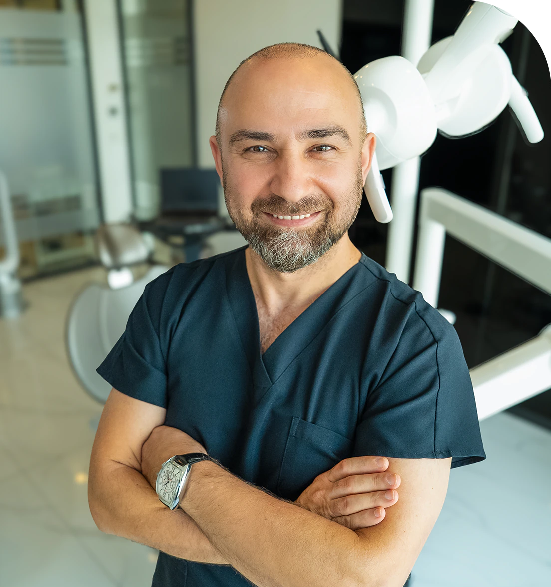
Dr. Dt. Ömer Atar provides care at his private clinic in Istanbul, combining his academic background and many years of clinical experience in dentistry. With a treatment approach that integrates aesthetic and functional solutions, he aims to achieve natural, balanced, and long-lasting results through personalized planning for each patient. Dr. Atar completed his education at the Marmara University Faculty of Dentistry, which he began in 1994, graduating in 2000. He continued his academic work in the Department of Prosthodontics at the same university. In 2007, he earned his PhD degree with his scientific thesis on implants and implant-supported prostheses in patients undergoing distraction osteogenesis. His academic expertise deepened in the fields of metal-free aesthetic systems, implant-supported prostheses, and cosmetic dentistry. Integrating modern technologies and digital dentistry into his practice, Dr. Ömer Atar achieves high success rates in treatments such as smile design, All-on-4/All-on-6 implant treatments, laminate veneers, zirconium crowns, teeth whitening, and gingival aesthetics (gummy smile). As a member of EDAD (Turkish Academy of Aesthetic Dentistry), he embraces a holistic approach focused on modern, functional, and natural outcomes in aesthetic dentistry.


