
 Türkçe
 Français
 Deutsch
 Nederlands
 Svenska

The clinical management of third molars (wisdom teeth) is a multidisciplinary treatment protocol that combines detailed radiological diagnosis, precise risk analysis, and minimally invasive surgical techniques. These procedures, which are the most commonly performed in oral surgery, involve not only the extraction of impacted teeth but also the preservation of the health of surrounding tissues and the prevention of pathological formations such as cysts. A successful treatment process begins with the accurate classification of the tooth’s position and root morphology, continues with techniques that respect the bone tissue during surgery, and is completed with the use of regenerative biomaterials that accelerate healing. This comprehensive approach enhances postoperative comfort while ensuring long-term oral and dental health.
Why Do Wisdom Teeth Remain Impacted and What Are the Criteria for Eruption?
Wisdom teeth generally attempt to erupt between the ages of 17 and 25. However, each individual’s genetic blueprint and jaw development are different. In males, these teeth usually begin to move between ages 20–23, and in females, between 21–22. The problem is that statistically, these are the most frequently impacted teeth in the jaw. When there is not enough space in the posterior region of the jawbone, the tooth either remains entirely within the bone or emerges partially through the gum in a “partially impacted” position.
The partially impacted position is one of the most concerning clinical situations. This is because a pocket that is impossible to clean forms between the piece of gum covering the tooth and the tooth itself. This pocket is a perfect trap for food debris and bacteria. While fully impacted teeth remain in a more sterile environment due to lack of contact with the outside world, partially impacted teeth are always at risk of infection. Therefore, the position of the tooth is vital in determining our clinical approach.
What Imaging Methods Do We Use During the Diagnostic Process?
Before deciding on a surgical intervention, we must perfectly map the area. Visual examination only reveals the tip of the iceberg. The shape of the roots, bone density, and most importantly, proximity to nerve canals can only be understood through radiological examinations.
Standard panoramic X-rays are our first resort. However, technical precision is very important here. Every detail—from the patient’s position to the angle of the device—affects the accuracy of the image. As clinicians, we want to see the end of the tooth root and how close it is to the nerve canal with millimetric accuracy.
If we suspect from the panoramic X-ray that the roots are touching the inferior alveolar nerve or if we see a pathological formation such as a cyst, two-dimensional images are insufficient. At this point, Cone Beam Computed Tomography (CBCT) comes into play. This technology allows us to obtain a three-dimensional model of the tooth and jawbone. Before starting the surgery, we know how much bone needs to be removed and whether the nerve passes on the right or left side of the tooth. This eliminates the risk of surprises.
How Is the Difficulty Level of Impacted Teeth Classified?
Patients sometimes ask, “My friend’s tooth was removed in 5 minutes, why do I need surgery?” The answer lies in the position and classification of the tooth. In surgical planning, we use scientific scales known as the “Pell & Gregory” and “Winter” classifications.
For example, we assess the amount of bone in front of the wisdom tooth. If there is enough room for the tooth to erupt, it is a simple procedure. However, if the tooth is completely embedded in the jawbone (ramus), it is classified as “Class 3” and requires advanced surgical techniques. The angulation of the tooth is also important; it may be vertical, horizontal, or inverted. Especially horizontally positioned and deeply embedded teeth require more bone removal, which may prolong the operation and increase the amount of postoperative swelling. Performing these analyses in advance is essential for properly informing the patient and managing expectations.
In Which Cases Is Extraction Absolutely Necessary?
The belief that every impacted tooth must be extracted is incorrect. If a tooth is completely within the bone, does not harm adjacent teeth, has not formed a cyst, and does not cause unexplained pain in the patient, we may prefer to monitor it. The main factor that leads us to a surgical decision is the presence or high risk of “pathology.”
However, in some cases, we cannot afford to wait, and extraction becomes inevitable. These are typically scenarios that compromise the patient’s quality of life or are confirmed to lead to more serious problems in the future.
Situations that absolutely require extraction include:
- Recurrent pericoronitis
- Uncontrollable tooth pain
- Extensive dental caries
- Root resorptions
- Cystic lesions
- Tumoral formations
- Advanced periodontal disease
- Temporomandibular joint disorders
Especially in lower wisdom teeth, the risk of pathology development is higher than in the upper jaw. Therefore, preventive extraction before problems arise is more widely accepted worldwide for lower impacted teeth.
Why Is Young Age Preferred for Surgery?
Patients usually tend to say, “I’m not in pain, I’ll wait.” However, from a surgical standpoint, the ideal time is not the 40s when complaints begin, but the early 20s when the tissues are still young. In young individuals, the jawbone is more flexible, which makes extraction technically easier. As age progresses, the bone becomes harder, the roots may fuse with the bone, and flexibility is lost.
Moreover, healing potential is much higher in young people. The tissue regeneration rate after surgery in a young patient is not the same as that of an older patient who may have systemic diseases (e.g., diabetes, hypertension). In extractions performed at an advanced age, the risk of jaw fracture and delayed nerve healing statistically increases. Therefore, if a tooth needs to be removed, postponing it does not benefit the patient; on the contrary, it complicates the process.
How Is Anesthesia Chosen and Is Pain Felt?
In modern dentistry, pain is a completely manageable issue. The biggest fear of our patients is feeling pain during the procedure. However, thanks to the powerful local anesthetics we use, nerve transmission in the area is completely stopped. The patient may feel pressure and touch, but not sharp pain.
In routine impacted tooth operations, local anesthesia is usually sufficient. However, in certain special cases, we consider sedation or general anesthesia to enhance patient comfort. Sedation is a highly comfortable method in which the patient is conscious but feels deep relaxation and sleepiness, and does not remember the procedure afterward.
Situations in which sedation or general anesthesia may be preferred include:
- Severe dental phobia
- Uncontrollable gag reflex
- Individuals with intellectual disabilities
- Allergy to local anesthesia
- Multiple and complex surgical procedures
Surgical Procedure Steps and How the Wisdom Tooth Is Removed
The surgical procedure is a systematic process that follows a series of rules. First, the area is anesthetized and sterilized. Then, a small, controlled incision is made in the gum to access the tooth and bone.
If the tooth is beneath the bone, a procedure called “osteotomy” is performed to remove the bone. This is the most critical stage of the surgery. Bone tissue is alive and very sensitive to heat. The frictional heat created by the rotary instruments used during bone removal can kill bone cells (necrosis). To prevent this, we surgeons perform the procedure while constantly cooling it with saline solution. Working at low speed and under plenty of irrigation is the most important factor in reducing postoperative pain and swelling.
Sometimes the tooth cannot be removed in one piece. To avoid damaging surrounding tissues and bone, segmenting the tooth and removing it in several pieces is a commonly used and safer method. After the tooth is removed, the remaining socket is cleaned, sharp bone edges are smoothed, and the wound is closed with sutures.
What Are CGF and Laser Techniques That Accelerate Healing?
In line with the “biological dentistry” approach adopted in our clinic, we do not simply extract the tooth and end the process. We use regenerative methods that trigger the body’s own healing mechanisms. Chief among these are CGF (Concentrated Growth Factor) and LLLT (Low-Level Laser Therapy).
Concentrated Growth Factor (CGF) Application:
This is a natural treatment carried out entirely with the patient’s own blood. A tube of blood is drawn from the patient before the operation and processed in a special centrifuge. This concentrates the healing cells (platelets) and growth factors in the blood. The resulting gel-like biological material is placed into the extraction socket.
Advantages of CGF application include:
- Accelerated tissue healing
- Less postoperative pain
- Control of bleeding
- Support for new bone formation
- Reduced risk of infection
Low-Level Laser Therapy (LLLT):
Laser technology provides “biostimulation” by stimulating the energy centers of cells. After the surgical procedure, the laser beam applied to the area accelerates tissue metabolism. This process is completely painless and takes only a few minutes.
Clinical benefits of laser therapy include:
- Suppression of swelling and edema
- Reduction of inflammation
- Pain relief
- Prevention of jaw opening limitations
What Should You Be Careful About After Surgery?
A successful surgery is the responsibility of the surgeon; however, a successful recovery process depends on patient compliance. Taking the antibiotics and painkillers prescribed after surgery on time is essential for infection and pain control.
One of the most important issues is the preservation of the “blood clot.” The clot formed in the extraction socket is the cornerstone of healing. If this clot becomes dislodged, the bone is exposed and severe pain begins.
Things to avoid during the healing process include:
- Hot food and drinks
- Hard and grainy foods
- Using a straw
- Smoking and alcohol consumption
- Vigorous mouth rinsing
- Touching the wound site
Applying ice (cold compress) externally to the area during the first 24 hours significantly prevents swelling on your face. From the next day onward, paying attention to oral hygiene, brushing gently, and using antiseptic mouthwashes will prevent the wound from becoming infected.
What Are the Possible Complications and How Are They Managed?
Every surgical intervention carries some risks. The most common situations after wisdom tooth operations are facial swelling (edema) and limited mouth opening (trismus). These are usually temporary and are the body’s natural response to trauma. The laser and CGF methods we use aim to minimize these complaints.
Sometimes, a condition known as “dry socket” (alveolitis) may develop. This occurs when the clot is disrupted and the bone is exposed, presenting as severe pain 2–3 days after the operation. In such a case, you should immediately consult your doctor; with a simple dressing, the pain can be controlled in a short time.
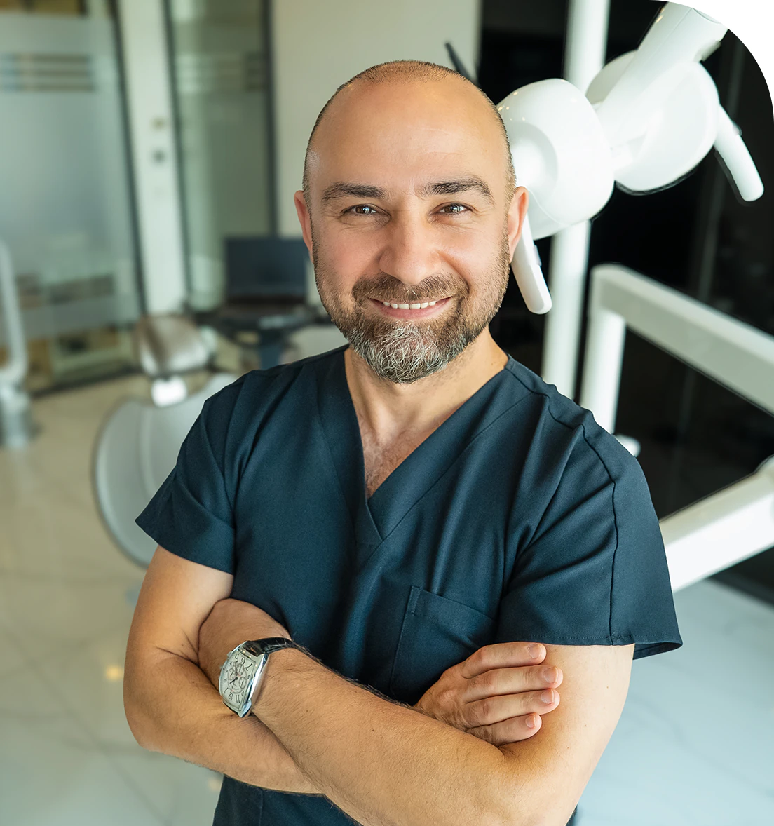
Dr. Dt. Ömer Atar provides care at his private clinic in Istanbul, combining his academic background and many years of clinical experience in dentistry. With a treatment approach that integrates aesthetic and functional solutions, he aims to achieve natural, balanced, and long-lasting results through personalized planning for each patient. Dr. Atar completed his education at the Marmara University Faculty of Dentistry, which he began in 1994, graduating in 2000. He continued his academic work in the Department of Prosthodontics at the same university. In 2007, he earned his PhD degree with his scientific thesis on implants and implant-supported prostheses in patients undergoing distraction osteogenesis. His academic expertise deepened in the fields of metal-free aesthetic systems, implant-supported prostheses, and cosmetic dentistry. Integrating modern technologies and digital dentistry into his practice, Dr. Ömer Atar achieves high success rates in treatments such as smile design, All-on-4/All-on-6 implant treatments, laminate veneers, zirconium crowns, teeth whitening, and gingival aesthetics (gummy smile). As a member of EDAD (Turkish Academy of Aesthetic Dentistry), he embraces a holistic approach focused on modern, functional, and natural outcomes in aesthetic dentistry.


