
 Türkçe
 Français
 Deutsch

An impacted tooth is a tooth that, although it has reached its eruption time, remains partially or completely within the jawbone or soft tissue. It is most commonly seen in wisdom teeth. Lack of space, incorrect tooth position, or developmental abnormalities can lead to the formation of impacted teeth.
Impacted teeth may cause pain, swelling, restricted jaw movements, and damage to adjacent teeth. They can also lead to complications such as cyst formation, infection, and gum inflammation. Some impacted teeth can remain in the mouth for years without causing any symptoms.
Treatment of impacted teeth is usually performed by surgical extraction. This procedure, carried out under local anesthesia by a dentist or oral surgeon, may vary depending on the tooth’s position. The post-operative healing process should be carefully monitored.
To reduce the risk of swelling, pain, and infection after surgery, antibiotics, painkillers, and good oral hygiene are recommended. Cold compresses and soft foods support healing. Individuals with impacted tooth problems should not neglect regular dental check-ups.
| WHAT YOU NEED TO KNOW | Information |
| Definition | An impacted tooth is one that is partially or completely trapped within the jawbone or gums, has not erupted in time, or cannot emerge due to lack of space. It is most commonly seen in wisdom teeth. |
| Most Commonly Impacted Teeth | Third molars (wisdom teeth), canine teeth (especially in the upper jaw), second premolars. |
| Causes | Lack of space in the gums or bone, genetic factors, blockage of the eruption path, misangled tooth development, failure of baby teeth to fall out on time. |
| Types | Fully impacted: The tooth remains completely within the bone. Partially impacted: Part of the tooth is visible in the mouth, while the other part is under the gums or bone. |
| Symptoms | Impacted teeth may sometimes show no symptoms. If symptoms exist: – Jaw pain, – Swollen gums, – Difficulty opening the mouth, – Bad breath, – Cavity or pressure sensation in the neighboring tooth. |
| Complications | Gum infection (pericoronitis), cyst formation, root resorption in neighboring teeth, damage to the jawbone, orthodontic issues. |
| Diagnostic Methods | The position, angle, and surrounding structures of the tooth are assessed using panoramic X-ray (OPG). If needed, 3D imaging (CBCT) is used. |
| Need for Treatment | Asymptomatic impacted teeth can be monitored. However, if there are risks such as infection, pain, cyst, or pressure, extraction is recommended. |
| Treatment Method | Surgical extraction of the impacted tooth (impacted tooth surgery). Performed under local anesthesia. General anesthesia may be preferred if necessary. |
| Surgical Stages | 1) The gum is cut to access the tooth, 2) If needed, bone is removed to expose the tooth, 3) The tooth may be sectioned and removed, 4) The area is cleaned and sutured. |
| Healing Process | Mild pain, swelling, and difficulty swallowing may occur for a few days after the operation. Healing is supported with ice application, prescribed medications, and soft foods. |
| Follow-up and Control | Sutures are removed after 7–10 days (if non-resorbable). Oral hygiene should be maintained, and smoking should be avoided. |
What is an Impacted Tooth?
An impacted tooth is one that remains partially or completely within the jawbone or under the gums despite the time for normal eruption having arrived. It is most frequently seen in wisdom teeth. Eruption may not occur due to lack of space, incorrect positioning, or genetic factors. Impacted teeth can sometimes cause issues such as pain, infection, or damage to adjacent teeth. In such cases, surgical intervention may be necessary.
Why Do Impacted Teeth Form and What Are the Causes?
The formation story of impacted teeth is actually quite complex and cannot be attributed to a single cause. Our patients often ask, “Why did my tooth remain impacted?” The answer usually starts with the evolution of human anatomy. In ancient times, people’s diets consisted of hard and raw foods, so their jaw structures were much larger and stronger. However, with modern humans consuming cooked and soft foods, the jawbone gradually became smaller. As the number of teeth remained the same while the jaw shrank, the last teeth to erupt—wisdom teeth—ran out of space.
However, lack of space is not the only reason. Sometimes genetic factors come into play. If either parent has a history of impacted teeth, the likelihood of occurrence in their child is quite high. Also, premature loss or failure to shed baby teeth in time can confuse the eruption path of the permanent tooth underneath, causing it to remain impacted. Some systemic conditions and syndromes can also slow down or completely halt the eruption mechanism of the teeth.
The main contributing factors that prepare the ground for impacted teeth formation are as follows:
- Lack of space in the jawbone
- Genetic predisposition
- Early loss of baby teeth
- Failure of baby tooth roots to resorb
- Presence of supernumerary (extra) teeth
- Cyst formations
- Presence of tumors
- Thick gingival tissue
- Hormonal imbalances
- Jaw trauma
- Infections
- Poor nutrition
What Are the Symptoms of an Impacted Tooth?
Impacted teeth don’t always make a noisy entrance. Sometimes, they can silently remain embedded in the bone for years and are only discovered incidentally during a routine X-ray examination. However, when they begin to move or become infected, the signals they send are quite pronounced. Patients often complain of a vague, radiating pain they cannot quite describe. This pain may not only occur at the site of the tooth but can also spread to the ear, temple, or neck.
This situation is especially different in partially impacted teeth. Part of the tooth is exposed in the mouth, while the other part remains covered by the gum. The gap between this piece of gum and the tooth becomes an ideal hiding spot for food debris and bacteria. Since this area is nearly impossible to clean, infections frequently occur. This condition is called “pericoronitis.” When an infection develops, the pain intensifies, swelling appears in the area, and the patient may even have difficulty opening their mouth.
Common signs indicating the presence of impacted teeth include:
- Redness in the gums
- Localized tenderness
- Pain in the jawbone
- Pain radiating to the ear
- Pain in the neck area
- Swelling in the lymph nodes
- Restricted mouth opening
- Difficulty swallowing
- Bad breath
- Unpleasant taste in the mouth
- Bleeding gums
- Facial asymmetry
What Are the Risks If Impacted Teeth Are Not Removed?
Many patients do not want their impacted teeth touched as long as there is no pain. However, pain is not the only reason we recommend surgical intervention. Impacted teeth can behave like a ticking time bomb inside the jawbone. One of the biggest risks is the pressure an impacted tooth applies to the healthy tooth in front of it as it tries to erupt. This pressure can cause root resorption of a healthy molar, which you are supposed to use for a lifetime, or lead to decay.
A more serious risk is the formation of cysts and tumors. The sac around the impacted tooth, known as the follicle, may undergo pathological changes over time and turn into a cystic structure. These cysts grow silently and gradually erode the jawbone from the inside. This not only weakens the bone but also makes the treatment process much more complex. In advanced cases of bone loss, the jawbone becomes so thin that it may face the risk of fracture from even a minor impact (pathologic fracture).
Complications that may arise from untreated impacted teeth include:
- Resorption of adjacent tooth roots
- Decay in the neighboring tooth
- Jaw cysts
- Jaw tumors
- Disruption of dental alignment
- Failure of orthodontic treatment
- Chronic infections
- Jawbone fractures
- Gum diseases
- Nerve compression
- Joint pain
What Are the Diagnostic and Imaging Methods for Impacted Teeth?
The foundation of proper treatment planning lies in an accurate diagnosis. Starting surgery without understanding the position of the impacted tooth, its depth within the bone, and its relation to surrounding tissues is like navigating in the dark. Therefore, imaging methods are always utilized during the initial examination. The most commonly used method in standard dental exams is the panoramic radiograph. This image allows us to see the entire upper and lower jaws, sinuses, joints, and impacted teeth in a single two-dimensional frame. For most cases, this imaging is sufficient.
However, there are situations where two-dimensional images do not reveal the whole picture. Especially when the roots of the lower wisdom teeth appear to overlap with the main nerve canal (mandibular canal) that provides sensation to the lip, or when the tooth is in a very complex position, more advanced imaging is required. This is where Cone Beam Computed Tomography (CBCT) comes in. Tomography provides a three-dimensional model of the tooth. Is the nerve passing in front of the tooth or behind it? Are the roots wrapping around the nerve? We get answers to these questions with millimetric precision.
Situations that necessitate CBCT imaging include:
- Close relationship with the nerve canal
- Suspected curvature in the roots
- Presence of cystic lesions
- Suspicion of tumors
- Root resorption of neighboring teeth
- Completely horizontal position
- Multiple impacted teeth
- Anatomical variations
How Is the Difficulty Level of an Impacted Tooth Surgery Determined?
Not all impacted tooth extractions are equally difficult, and not every operation takes the same amount of time. As dentists, when evaluating radiographic images, we mentally assess the surgical difficulty of that tooth using specific scientific classifications (such as Winter and Pell & Gregory). This analysis guides us when informing the patient about the procedure and estimating the recovery process.
The angle of the tooth is one of the most important factors influencing the surgery. For example, extracting a tooth slightly inclined forward is usually easier, whereas removing a tooth tilted backward or fully inverted requires more technical and precise work. Similarly, the depth of the tooth matters. If part of the tooth is visible in the mouth, the procedure may be simpler, but if it is completely embedded in the bone and covered with a thick layer of bone, removal of that bone may be necessary, prolonging the operation.
Key factors that make the surgery more difficult include:
- Tooth being fully embedded in the bone
- Backward-tilted position
- Horizontal orientation
- Long and thin roots
- Hook-shaped roots
- Roots in contact with the nerve
- Advanced age
- Loss of bone elasticity
- Limited mouth opening
- Strong gag reflex
- Large tongue
What Is the Treatment Approach for Canine Teeth?
When talking about wisdom teeth, the focus is usually on “extraction” because their contribution to chewing is limited, and their absence is not a major loss. However, when it comes to impacted canine teeth, our approach changes completely. Canines are located at the corners of the mouth, play a major role in facial aesthetics, and have the longest and strongest roots. Therefore, extracting an impacted canine is the last and least desired option for us.
Our primary goal with canines is to save the tooth and bring it to its rightful place in the dental arch. This process requires teamwork. We usually collaborate with orthodontists. Through a surgical procedure, we uncover the tooth and attach a small appliance (bracket) to it. Then, using braces, we gradually pull the tooth downward over the course of several months. However, if the tooth is in a severely abnormal position, causing damage to adjacent roots, or is immobile (ankylosed), extraction may become necessary.
Treatment options for canine teeth include:
- Orthodontic traction
- Surgical exposure
- Transplantation
- Surgical extraction
- Implant treatment
- Bridge prosthesis
- Monitoring under observation
What Are the Stages of Impacted Tooth Surgery?
Although the word “surgery” may frighten patients, today these procedures are quite routine, painless, and comfortable. Thanks to advances in technology and anesthetic agents, patients feel no pain while sitting in the chair. The procedure begins in full compliance with sterilization protocols.
The process starts with the administration of strong local anesthesia. We wait until the area is completely numb. From this point, the patient only feels touch and pressure, not sharp pain. A small incision is made in the gum to access the tooth. If the tooth is under bone, the overlying bone tissue is gently removed using special tools and plenty of irrigation.
The most important step is the removal of the tooth. Trying to extract large and deeply impacted teeth in one piece requires significant bone removal and puts excessive pressure on the jawbone. Therefore, we usually prefer to section the tooth into several parts (a process called sectioning) before removal. This “divide and conquer” method reduces trauma and speeds up healing. Finally, the area is cleaned and closed with sutures.
Basic steps of the surgical procedure include:
- Application of local anesthesia
- Incision of the gum
- Elevation of the gum tissue
- Removal of bone tissue
- Sectioning of the tooth
- Removal of the sections
- Cleaning of cyst or follicle
- Smoothing of bone edges
- Irrigation of the area
- Suturing
- Bleeding control
What Is the Coronectomy Method and Who Is It For?
Sometimes, radiographs or CT scans show that the tooth roots are dangerously close to the nerve canal. In such cases, attempting full extraction may damage the nerve and cause temporary or permanent numbness in the lip. Following the principle of “Do no harm,” we apply a special method called “Coronectomy” in these high-risk cases.
In a coronectomy, the entire tooth is not removed. Only the crown portion—the part that causes pain and infection—is cut and extracted. The root portion, which is in contact with the nerve, is left undisturbed within the bone. The remaining root surface is smoothed, and the area is completely closed with gum tissue. This method eliminates the risk of nerve damage. The remaining root piece usually fuses with the bone over time and typically causes no problems for life. This technique is a lifesaving risk-management tool.
Advantages of the coronectomy procedure include:
- Prevention of nerve damage
- Avoidance of lip numbness
- Preservation of the jawbone
- Less traumatic surgery
- Faster healing process
- Reduced risk of complications
- Lower risk of jaw fracture
What Is the Postoperative Healing Process and What Should Be Considered?
A successful operation is only half the battle; the other half is the healing period spent at home. After the surgery, it is biologically normal for the body to react with swelling or mild pain in the area. Slight bleeding may occur on the first day. During this time, patients should remain calm and strictly follow the dentist’s instructions.
Swelling (edema), in particular, is part of the body’s healing response. The golden rule to minimize swelling is cold application (ice compress). Applying ice externally for 20 minutes on and 20 minutes off during the first 24 hours after surgery significantly reduces both swelling and pain. Oral hygiene is also critical; however, brushing or vigorous rinsing on the first day may disrupt the blood clot.
Vital post-procedure rules include:
- Bite down on the gauze for 45 minutes
- Do not spit
- Do not use a straw
- Do not smoke
- Do not consume alcohol
- Avoid hot foods
- Avoid foods with particles or seeds
- Take painkillers regularly
- Keep your head elevated
- Avoid strenuous exercise
- Apply ice
- Use prescribed medications
How Should Nutrition Be Managed During the Healing Process?
One of the most challenging aspects for patients after impacted tooth surgery is nutrition. Limited jaw movement and difficulty swallowing can make eating more difficult. However, the body needs energy and protein to heal. In the first 24 hours, extremely hot foods should be strictly avoided; heat can dilate blood vessels and restart bleeding. Warm or cold foods are preferable.
Hard, chewy, shelled, or seedy foods (such as rice, sesame, strawberries) can enter the surgical wound site and cause infection. Therefore, a “soft diet” should be followed for the first few days. Foods that require no chewing and have a puree-like consistency are both comfortable and safe. Drinking plenty of water is essential for healing, but using a straw creates a vacuum effect that can dislodge the protective blood clot at the surgical site and lead to a painful condition known as “dry socket.”
Recommended foods include:
- Warm soups
- Yogurt and ayran (diluted yogurt drink)
- Mashed potatoes
- Pudding
- Fruit purees
- Omelet
- Ice cream
- Pasta
- Boiled vegetables
- Smoothie drinks
Frequently Asked Questions
What is an impacted tooth and which teeth are most commonly affected?
An impacted tooth is one that is partially or completely unable to emerge from the jawbone. It is most commonly seen in third molars, i.e., wisdom teeth, but canine and molar teeth can also be impacted.
What causes an impacted tooth?
Narrow jaws, genetic factors, failure of baby teeth to fall out on time, and the position of neighboring teeth can lead to impaction. Sometimes, even if a tooth is fully developed, it may not have enough space to erupt.
What are the symptoms of an impacted tooth?
Symptoms may include jaw pain, difficulty opening the mouth, swelling in the gums, bad breath, and sometimes pain radiating to the ear. However, some impacted teeth may not show symptoms for a long time.
Can an impacted tooth damage nearby teeth?
Yes, an impacted tooth can exert pressure on the roots of adjacent teeth, causing pain, root resorption, or even tooth loss. It also carries a risk of cyst formation.
What happens if an impacted tooth becomes infected?
If infection develops around an impacted tooth, it leads to a condition called pericoronitis. Symptoms may include severe pain, swelling, fever, and limited jaw movement.
How is an impacted tooth diagnosed?
It can be detected during a routine dental examination. The position, development, and effect on surrounding tissues are evaluated in detail using panoramic X-rays or dental CT scans.
Should every impacted tooth be extracted?
Not every impacted tooth needs to be removed. However, if it causes pain, infection, cyst formation, or damage to nearby teeth, surgical removal is recommended.
How is impacted tooth surgery performed?
Under local anesthesia, surgical incisions are made to access the gum and jawbone. The tooth may be sectioned and removed in parts. The procedure typically takes 20–45 minutes and is completed with sutures.
What is the recovery process like after impacted tooth extraction?
Recovery generally takes 7–10 days. Swelling, pain, and mild bleeding may occur in the first few days. The process is managed with antibiotics and painkillers. Cold compresses and soft foods are advised.
What complications can arise if an impacted tooth is not removed?
Serious problems such as cyst formation, decay and root resorption in adjacent teeth, gum infection, chronic pain, and, rarely, jawbone weakening may occur. Early intervention is important.
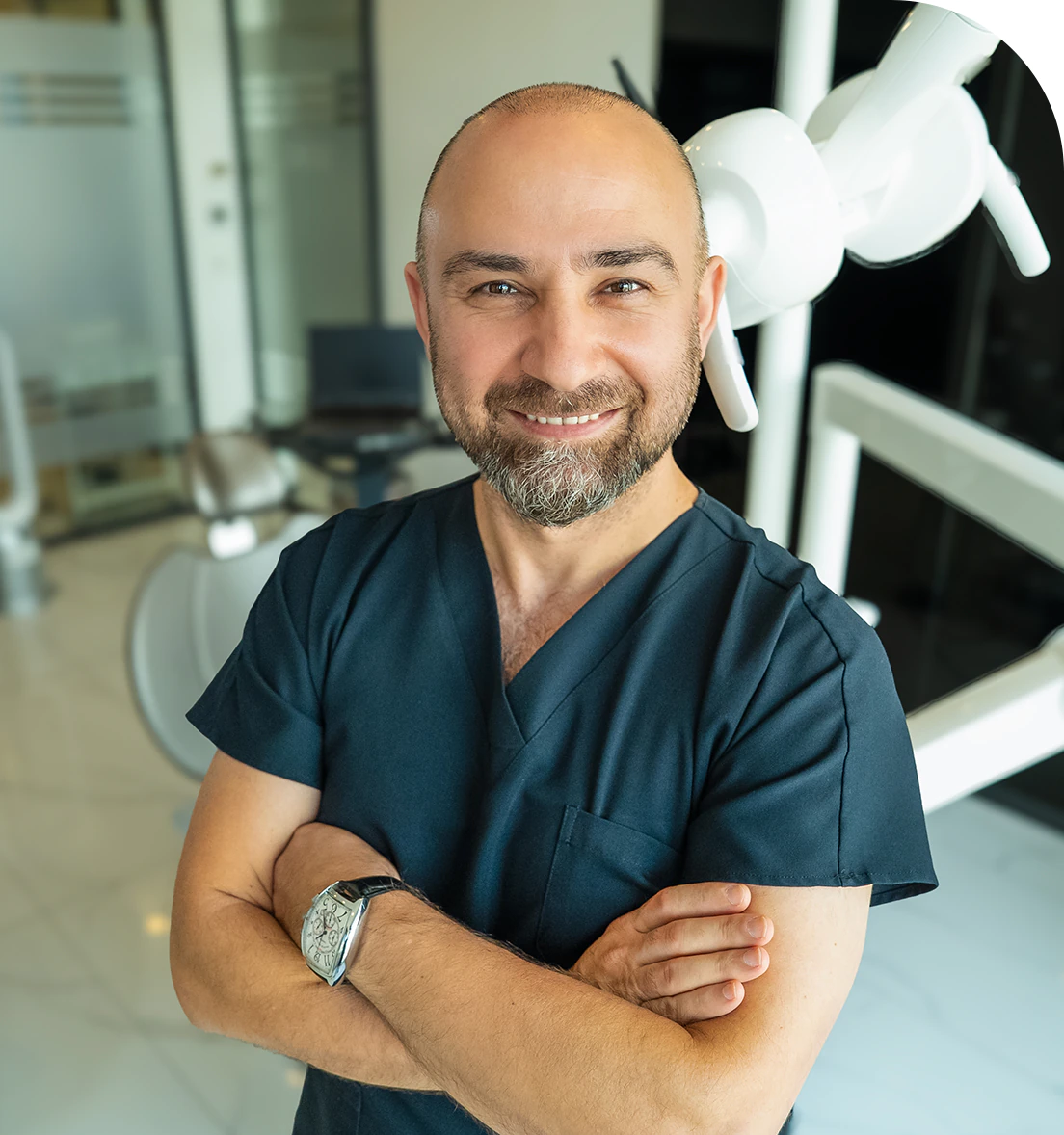
Dr. Dt. Ömer Atar provides care at his private clinic in Istanbul, combining his academic background and many years of clinical experience in dentistry. With a treatment approach that integrates aesthetic and functional solutions, he aims to achieve natural, balanced, and long-lasting results through personalized planning for each patient. Dr. Atar completed his education at the Marmara University Faculty of Dentistry, which he began in 1994, graduating in 2000. He continued his academic work in the Department of Prosthodontics at the same university. In 2007, he earned his PhD degree with his scientific thesis on implants and implant-supported prostheses in patients undergoing distraction osteogenesis. His academic expertise deepened in the fields of metal-free aesthetic systems, implant-supported prostheses, and cosmetic dentistry. Integrating modern technologies and digital dentistry into his practice, Dr. Ömer Atar achieves high success rates in treatments such as smile design, All-on-4/All-on-6 implant treatments, laminate veneers, zirconium crowns, teeth whitening, and gingival aesthetics (gummy smile). As a member of EDAD (Turkish Academy of Aesthetic Dentistry), he embraces a holistic approach focused on modern, functional, and natural outcomes in aesthetic dentistry.


