
 Türkçe
 Français
 Deutsch
 Nederlands
 Svenska

Molars, also known as posterior teeth, are permanent teeth located at the very back of the oral cavity. They possess broad chewing surfaces and strong root structures and are primarily responsible for grinding food. With three on each half of the jaw — totaling twelve in the mouth — they mark the starting point of the digestive system while also supporting temporomandibular joint health and maintaining the lower third facial height. Due to their resistance to high chewing forces and complex anatomy, molars are fundamental pillars of oral health that require advanced diagnostic techniques and precise treatment approaches.
How does the root structure of molars affect treatment?
You can think of molars as the “millstones” of our mouths. To withstand such a heavy workload, they have multiple roots, much like trees that anchor deep into the ground. Typically, mandibular molars have two roots, while maxillary molars have three. But this is only the visible and commonly known part. What truly challenges us and determines the success of the treatment is the number and shape of the canals within those roots.
Maxillary molars, in particular, may have palatal roots that curve like a hook instead of descending straight. These curves can make it difficult for the thin, needle-like instruments (files) used in root canal therapy to progress. Even more interestingly, mandibular molars can sometimes have an “extra” root or an unexpected canal. These additional anatomical features, known in medical literature as “Radix Entomolaris,” can be so hidden that they are easily missed during treatment. If a root canal is being performed and the clinician fails to identify and clean this hidden chamber, the infection within the tooth will persist. The patient may believe the treatment was successful, only to experience an abscess at the root of the same tooth months or years later. That’s why molar treatment demands significantly more patience, experience, and anatomical knowledge than anterior teeth.
Are X-rays sufficient to detect every issue?
Periapical radiographs — the small X-ray films we’ve used in dentistry for years — are essential diagnostic tools. However, when it comes to molars, these images sometimes only reveal part of the truth. Standard X-rays are two-dimensional and lack depth. When taking an image of a three-rooted molar, one root may obscure another, or a cyst hidden within the bone may be concealed by root shadows.
This situation is similar to an iceberg where the bulk remains unseen beneath the surface. Especially in cases that require retreatment of root canals or in unexplained pain scenarios, two-dimensional images are insufficient. At this point, Cone Beam Computed Tomography (CBCT) — one of the most powerful technologies available — comes into play. With these devices, we can examine the tooth layer by layer in 3D. We can see exactly where the root curves, where the canal bifurcates, or how far an infection has spread near the root apex, all with millimetric accuracy. This mapping minimizes the risks of root perforation or underfilled canals during treatment.
Why do pain and sensitivity occur if there’s no cavity?
One of the most frequently reported and diagnostically challenging conditions we encounter is “Cracked Tooth Syndrome.” Patients often say there is no visible decay and they are not particularly sensitive to hot or cold stimuli. Yet their complaint is very specific: “When I chew — especially on something hard — I get a sharp, lightning-like pain, and the moment I stop biting, the pain disappears.” This is the hallmark of a microfracture that is not visible but has compromised the structural integrity of the tooth.
Over the years, molars endure tons of pressure, leading to fatigue in the enamel. When biting down, the microscopic crack may slightly open, and when the pressure is released, it snaps shut. This movement causes fluid displacement in the pulp tissue, triggering an acute pain response. If not treated early, the crack may deepen into the nerve or even split the tooth vertically, necessitating extraction.
Common triggers for this syndrome include:
- Bruxism (teeth clenching at night)
- Teeth grinding
- Chewing ice
- Cracking hard candy
- Biting olive pits
- Large, old fillings
- Deep fissures in teeth
- Sharp cusp structures
- Traumatic injuries
Why choose porcelain onlays over large fillings?
When a deep cavity is cleaned out or an old, large metal filling is removed in a molar, the remaining healthy tooth structure can be very thin and fragile. Filling this cavity with standard materials (composite or amalgam) may do more harm than good. Under chewing forces, a large filling mass can act like a wedge and push the weakened tooth walls outward, eventually causing the tooth to fracture at its weakest point.
To prevent this, we prefer porcelain restorations called “onlays” that are fabricated in a laboratory. You can think of them as custom-made puzzle pieces that complete the missing part of the tooth. Onlays do not behave like a filling packed into the tooth; instead, they cover and reinforce the weakened walls from above, locking them together — much like the metal hoops around a barrel. Onlays are aesthetically matched to the natural tooth color and are biomechanically the safest solution for protecting the tooth. In teeth at risk of cracked tooth syndrome, this approach — which preserves the tooth by only repairing the damaged portion instead of fully crowning it — is a cornerstone of conservative dentistry.
What can be done to prevent cavities in children?
Molars begin to erupt in children around the age of 6. These are the first permanent teeth and, unfortunately, the ones most prone to decay. This is because their chewing surfaces are not flat; instead, they contain deep grooves, pits, and fissures. These fissures can be so narrow that even the bristles of the best toothbrushes can’t reach into them, whereas bacteria and sugary food particles can easily settle there and begin decaying the enamel from within.
The most effective method to prevent this in children is the application of “fissure sealants.” This procedure is completely painless and requires no drilling. The deep grooves are simply sealed with a protective, flowable resin that creates a smooth and brushable surface.
Preventive measures and recommendations include:
- Fissure sealant application
- Topical fluoride varnish
- Regular tooth brushing
- Limiting sugary foods
- Routine dental checkups
- Healthy dietary habits
- Saliva (moisture) control
Should impacted wisdom teeth always be removed?
Wisdom teeth — the third molars — are increasingly struggling to find room in the modern human jaw. Patients often ask, “I have no pain; should I still have it removed?” For us dentists, the decision to extract a wisdom tooth is based not just on current symptoms but also on the potential future damage it may cause.
A completely or partially impacted wisdom tooth is like a ticking time bomb. The greatest risk is not to itself, but to the healthy second molar in front of it — a tooth you need for life. A partially erupted wisdom tooth creates a pocket in the gum that cannot be brushed, making it a trap for food debris and a breeding ground for infections (pericoronitis) and cysts. Worse, a horizontally positioned wisdom tooth can press against the adjacent tooth’s root, leading to decay or resorption of that otherwise healthy tooth. Therefore, the most prudent approach is to remove high-risk wisdom teeth in young patients to protect adjacent teeth.
Factors influencing the extraction decision include:
- Recurrent infections
- Advanced decay
- Risk of cyst or tumor formation
- Damage to neighboring teeth
- Need for orthodontic treatment
- Prosthetic planning
- Chronic cheek biting
What’s the best solution to replace a lost molar?
Despite all efforts, when a molar is lost, the gap must be filled. Nature abhors a vacuum — when a tooth is extracted, the adjacent tooth begins to tilt forward, and the opposing tooth starts to over-erupt into the empty space. This disrupts the entire balance of the jaw.
In the past, these gaps were filled using bridge prostheses. However, to place a bridge, the healthy teeth on either side had to be ground down — a method no longer favored from a biological standpoint. Today, “single-tooth implants” provide a definitive solution. An implant is a titanium screw that mimics the root of the lost tooth and is placed only in the vacant area, leaving neighboring teeth untouched.
However, when placing implants in the molar region, we must also pay close attention to one critical factor: gum health. The gum surrounding the implant must be firm, resilient, and pink — what we call “keratinized gingiva.” If this tissue is insufficient, the risk of bacterial accumulation and infection increases. Therefore, we may perform minor soft tissue procedures during implant surgery to ensure long-term success.
Advantages of implant treatment include:
- Preservation of adjacent teeth
- Prevention of jawbone resorption
- Natural chewing sensation
- Long-term durability
- High success rate
- Ease of cleaning
- Esthetic appearance
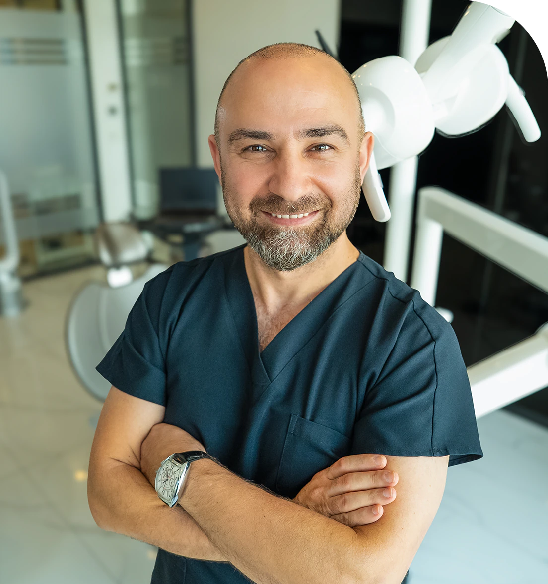
Dr. Dt. Ömer Atar provides care at his private clinic in Istanbul, combining his academic background and many years of clinical experience in dentistry. With a treatment approach that integrates aesthetic and functional solutions, he aims to achieve natural, balanced, and long-lasting results through personalized planning for each patient. Dr. Atar completed his education at the Marmara University Faculty of Dentistry, which he began in 1994, graduating in 2000. He continued his academic work in the Department of Prosthodontics at the same university. In 2007, he earned his PhD degree with his scientific thesis on implants and implant-supported prostheses in patients undergoing distraction osteogenesis. His academic expertise deepened in the fields of metal-free aesthetic systems, implant-supported prostheses, and cosmetic dentistry. Integrating modern technologies and digital dentistry into his practice, Dr. Ömer Atar achieves high success rates in treatments such as smile design, All-on-4/All-on-6 implant treatments, laminate veneers, zirconium crowns, teeth whitening, and gingival aesthetics (gummy smile). As a member of EDAD (Turkish Academy of Aesthetic Dentistry), he embraces a holistic approach focused on modern, functional, and natural outcomes in aesthetic dentistry.


